3Q6P | pdb_00003q6p

Salivary protein from Lutzomyia longipalpis. Selenomethionine derivative


Experimental Data Snapshot

  • Method: X-RAY DIFFRACTION
  • Resolution: 2.75 Å
  • R-Value Free: 
    0.261 (Depositor), 0.261 (DCC) 
  • R-Value Work: 
    0.197 (Depositor), 0.196 (DCC) 
  • R-Value Observed: 
    0.200 (Depositor) 

wwPDB Validation 3D Report Full Report

Validation slider image for 3Q6P

This is version 1.3 of the entry. See complete history

Literature

Structure and function of a "yellow" protein from saliva of the sand fly Lutzomyia longipalpis that confers protective immunity against Leishmania major infection.

Xu, X.Oliveira, F.Chang, B.W.Collin, N.Gomes, R.Teixeira, C.Reynoso, D.My Pham, V.Elnaiem, D.E.Kamhawi, S.Ribeiro, J.M.Valenzuela, J.G.Andersen, J.F.

(2011) J Biological Chem 286: 32383-32393

  • DOI: https://doi.org/10.1074/jbc.M111.268904
  • Primary Citation Related Structures: 
    3Q6K, 3Q6P, 3Q6T

  • PubMed Abstract: 

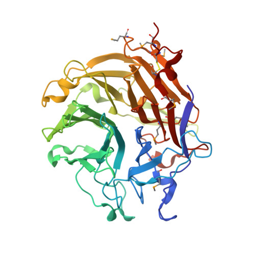
    LJM11, an abundant salivary protein from the sand fly Lutzomyia longipalpis, belongs to the insect "yellow" family of proteins. In this study, we immunized mice with 17 plasmids encoding L. longiplapis salivary proteins and demonstrated that LJM11 confers protective immunity against Leishmania major infection. This protection correlates with a strong induction of a delayed type hypersensitivity (DTH) response following exposure to L. longipalpis saliva. Additionally, splenocytes of exposed mice produce IFN-γ upon stimulation with LJM11, demonstrating the systemic induction of Th1 immunity by this protein. In contrast to LJM11, LJM111, another yellow protein from L. longipalpis saliva, does not produce a DTH response in these mice, suggesting that structural or functional features specific to LJM11 are important for the induction of a robust DTH response. To examine these features, we used calorimetric analysis to probe a possible ligand binding function for the salivary yellow proteins. LJM11, LJM111, and LJM17 all acted as high affinity binders of prohemostatic and proinflammatory biogenic amines, particularly serotonin, catecholamines, and histamine. We also determined the crystal structure of LJM11, revealing a six-bladed β-propeller fold with a single ligand binding pocket located in the central part of the propeller structure on one face of the molecule. A hypothetical model of LJM11 suggests a positive electrostatic potential on the face containing entry to the ligand binding pocket, whereas LJM111 is negative to neutral over its entire surface. This may be the reason for differences in antigenicity between the two proteins.


  • Organizational Affiliation
    • Laboratory of Malaria and Vector Research, NIAID, National Institutes of Health, Rockville, Maryland 20852, USA.

Macromolecule Content 

  • Total Structure Weight: 87.46 kDa 
  • Atom Count: 6,190 
  • Modeled Residue Count: 762 
  • Deposited Residue Count: 762 
  • Unique protein chains: 1

Macromolecules

Find similar proteins by:|  3D Structure
Entity ID: 1
MoleculeChains  Sequence LengthOrganismDetailsImage
43.2 kDa salivary protein
A, B
381Lutzomyia longipalpisMutation(s): 0 
Gene Names: LJM-11LJM11_Clu9
UniProt
Find proteins for Q5WPU9 (Lutzomyia longipalpis)
Explore Q5WPU9 
Go to UniProtKB:  Q5WPU9
Entity Groups
Sequence Clusters30% Identity50% Identity70% Identity90% Identity95% Identity100% Identity
UniProt GroupQ5WPU9
Sequence Annotations
Expand
Reference Sequence

Small Molecules

Ligands 1 Unique
IDChains Name / Formula / InChI Key2D Diagram3D Interactions
CIT

Query on CIT


Download:Ideal Coordinates CCD File
C [auth B]CITRIC ACID
C6 H8 O7
KRKNYBCHXYNGOX-UHFFFAOYSA-N
Modified Residues  1 Unique
IDChains TypeFormula2D DiagramParent
MSE
Query on MSE
A, B
L-PEPTIDE LINKINGC5 H11 N O2 SeMET

Experimental Data & Validation

Experimental Data

  • Method: X-RAY DIFFRACTION
  • Resolution: 2.75 Å
  • R-Value Free:  0.261 (Depositor), 0.261 (DCC) 
  • R-Value Work:  0.197 (Depositor), 0.196 (DCC) 
  • R-Value Observed: 0.200 (Depositor) 
Space Group: P 62 2 2
Unit Cell:
Length ( Å )Angle ( ˚ )
a = 120.355α = 90
b = 120.355β = 90
c = 248.934γ = 120
Software Package:
Software NamePurpose
DENZOdata reduction
SCALEPACKdata scaling
SHELXphasing
REFMACrefinement
PDB_EXTRACTdata extraction
SHELXDphasing
SHELXEmodel building

Structure Validation

View Full Validation Report


Entry History 

Deposition Data

Revision History  (Full details and data files)

  • Version 1.0: 2011-07-27
    Type: Initial release
  • Version 1.1: 2017-11-08
    Changes: Refinement description
  • Version 1.2: 2019-10-23
    Changes: Data collection, Database references, Derived calculations
  • Version 1.3: 2024-11-27
    Changes: Data collection, Database references, Derived calculations, Structure summary