3Q2M

Crystal Structure of Spectinomycin Phosphotransferase, APH(9)-Ia, Protein Kinase Inhibitor CKI-7 Complex


Experimental Data Snapshot

  • Method: X-RAY DIFFRACTION
  • Resolution: 2.90 Å
  • R-Value Free: 0.279 
  • R-Value Work: 0.221 
  • R-Value Observed: 0.221 

Starting Model: experimental
View more details

wwPDB Validation   3D Report Full Report


Ligand Structure Quality Assessment 


This is version 1.3 of the entry. See complete history


Literature

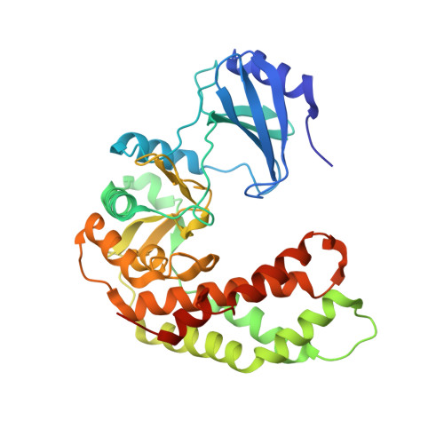
Crystal structures of two aminoglycoside kinases bound with a eukaryotic protein kinase inhibitor.

Fong, D.H.Xiong, B.Hwang, J.Berghuis, A.M.

(2011) PLoS One 6: e19589-e19589

  • DOI: https://doi.org/10.1371/journal.pone.0019589
  • Primary Citation of Related Structures:  
    3Q2J, 3Q2M

  • PubMed Abstract: 

    Antibiotic resistance is recognized as a growing healthcare problem. To address this issue, one strategy is to thwart the causal mechanism using an adjuvant in partner with the antibiotic. Aminoglycosides are a class of clinically important antibiotics used for the treatment of serious infections. Their usefulness has been compromised predominantly due to drug inactivation by aminoglycoside-modifying enzymes, such as aminoglycoside phosphotransferases or kinases. These kinases are structurally homologous to eukaryotic Ser/Thr and Tyr protein kinases and it has been shown that some can be inhibited by select protein kinase inhibitors. The aminoglycoside kinase, APH(3')-IIIa, can be inhibited by CKI-7, an ATP-competitive inhibitor for the casein kinase 1. We have determined that CKI-7 is also a moderate inhibitor for the atypical APH(9)-Ia. Here we present the crystal structures of CKI-7-bound APH(3')-IIIa and APH(9)-Ia, the first structures of a eukaryotic protein kinase inhibitor in complex with bacterial kinases. CKI-7 binds to the nucleotide-binding pocket of the enzymes and its binding alters the conformation of the nucleotide-binding loop, the segment homologous to the glycine-rich loop in eukaryotic protein kinases. Comparison of these structures with the CKI-7-bound casein kinase 1 reveals features in the binding pockets that are distinct in the bacterial kinases and could be exploited for the design of a bacterial kinase specific inhibitor. Our results provide evidence that an inhibitor for a subset of APHs can be developed in order to curtail resistance to aminoglycosides.


  • Organizational Affiliation

    Department of Biochemistry, McGill University, Montreal, Quebec, Canada.


Macromolecules
Find similar proteins by:  (by identity cutoff)  |  3D Structure
Entity ID: 1
MoleculeChains Sequence LengthOrganismDetailsImage
Spectinomycin phosphotransferase339Legionella pneumophila 130bMutation(s): 0 
Gene Names: aphLPW_21961
UniProt
Find proteins for O06916 (Legionella pneumophila)
Explore O06916 
Go to UniProtKB:  O06916
Entity Groups  
Sequence Clusters30% Identity50% Identity70% Identity90% Identity95% Identity100% Identity
UniProt GroupO06916
Sequence Annotations
Expand
  • Reference Sequence
Small Molecules
Ligands 2 Unique
IDChains Name / Formula / InChI Key2D Diagram3D Interactions
CKI
Query on CKI

Download Ideal Coordinates CCD File 
B [auth A]N-(2-AMINOETHYL)-5-CHLOROISOQUINOLINE-8-SULFONAMIDE
C11 H12 Cl N3 O2 S
OGKYMFFYOWUTKV-UHFFFAOYSA-N
NI
Query on NI

Download Ideal Coordinates CCD File 
C [auth A]NICKEL (II) ION
Ni
VEQPNABPJHWNSG-UHFFFAOYSA-N
Binding Affinity Annotations 
IDSourceBinding Affinity
CKI PDBBind:  3Q2M Ki: 1.59e+5 (nM) from 1 assay(s)
Experimental Data & Validation

Experimental Data

  • Method: X-RAY DIFFRACTION
  • Resolution: 2.90 Å
  • R-Value Free: 0.279 
  • R-Value Work: 0.221 
  • R-Value Observed: 0.221 
  • Space Group: P 31 2 1
Unit Cell:
Length ( Å )Angle ( ˚ )
a = 74.3α = 90
b = 74.3β = 90
c = 137.024γ = 120
Software Package:
Software NamePurpose
CrystalCleardata collection
PHASERphasing
CNSrefinement
HKL-2000data reduction
HKL-2000data scaling

Structure Validation

View Full Validation Report


Ligand Structure Quality Assessment 


Entry History 

Deposition Data

Revision History  (Full details and data files)

  • Version 1.0: 2011-05-18
    Type: Initial release
  • Version 1.1: 2011-07-13
    Changes: Version format compliance
  • Version 1.2: 2013-06-19
    Changes: Database references
  • Version 1.3: 2023-09-13
    Changes: Data collection, Database references, Derived calculations, Refinement description