3PZF | pdb_00003pzf

1.75A resolution structure of Serpin-2 from Anopheles gambiae


Experimental Data Snapshot

  • Method: X-RAY DIFFRACTION
  • Resolution: 1.75 Å
  • R-Value Free: 
    0.207 (Depositor), 0.209 (DCC) 
  • R-Value Work: 
    0.173 (Depositor), 0.176 (DCC) 
  • R-Value Observed: 
    0.175 (Depositor) 

Starting Model: experimental
View more details

wwPDB Validation 3D Report Full Report

Validation slider image for 3PZF

This is version 1.5 of the entry. See complete history

Literature

Crystal structure of native Anopheles gambiae serpin-2, a negative regulator of melanization in mosquitoes.

An, C.Lovell, S.Kanost, M.R.Battaile, K.P.Michel, K.

(2011) Proteins 79: 1999-2003

  • DOI: https://doi.org/10.1002/prot.23002
  • Primary Citation Related Structures: 
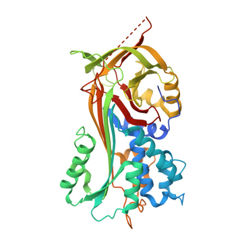
    3PZF

  • PubMed Abstract: 

    Serpins are the dominant group of protease inhibitors in metazoans that control a wide variety of biological processes including major innate immune reactions. One of these inhibitors, SRPN2, controls melanization in mosquitoes – a powerful, arthropod-specific innate immune response. SRPN2 depletion from the hemolymph of adult female mosquitoes significantly reduces longevity and therefore this serpin is a potential target for novel insecticides. We report here the crystal structure of SRPN2 in its native conformation from the African malaria mosquito, Anopheles gambiae to 1.75 Å resolution. SRPN2 adopts a similar fold as observed for other serpins with a core of three β-sheets surrounded by nine α-helices with an exposed reactive center loop (RCL) that extends from the protein body. Similar to other native serpin structures, several residues within the reactive center loop were disordered and could not be modeled. Intriguingly, the N-terminal hinge of the RCL in SRPN2 was found to be inserted into β-sheet A, suggesting a potential activation mechanism analogous to heparin-mediated activation of Antithrombin III.


  • Organizational Affiliation
    • Division of Biology, Kansas State University, Manhattan, Kansas 66506, USA.

Macromolecule Content 

  • Total Structure Weight: 45.36 kDa 
  • Atom Count: 3,118 
  • Modeled Residue Count: 369 
  • Deposited Residue Count: 397 
  • Unique protein chains: 1

Macromolecules

Find similar proteins by:|  3D Structure
Entity ID: 1
MoleculeChains  Sequence LengthOrganismDetailsImage
Serpin 2397Anopheles gambiaeMutation(s): 0 
Gene Names: SRPN2
UniProt
Find proteins for Q7QIJ8 (Anopheles gambiae)
Explore Q7QIJ8 
Go to UniProtKB:  Q7QIJ8
Entity Groups
Sequence Clusters30% Identity50% Identity70% Identity90% Identity95% Identity100% Identity
UniProt GroupQ7QIJ8
Sequence Annotations
Expand
Reference Sequence

Experimental Data & Validation

Experimental Data

  • Method: X-RAY DIFFRACTION
  • Resolution: 1.75 Å
  • R-Value Free:  0.207 (Depositor), 0.209 (DCC) 
  • R-Value Work:  0.173 (Depositor), 0.176 (DCC) 
  • R-Value Observed: 0.175 (Depositor) 
Space Group: P 63
Unit Cell:
Length ( Å )Angle ( ˚ )
a = 96.538α = 90
b = 96.538β = 90
c = 78.289γ = 120
Software Package:
Software NamePurpose
PHENIXrefinement
PHASERphasing
REFMACrefinement
PDB_EXTRACTdata extraction
DENZOdata reduction
SCALEPACKdata scaling

Structure Validation

View Full Validation Report


Entry History 

Deposition Data

Revision History  (Full details and data files)

  • Version 1.0: 2011-02-16
    Type: Initial release
  • Version 1.1: 2011-07-13
    Changes: Version format compliance
  • Version 1.2: 2017-11-08
    Changes: Refinement description
  • Version 1.3: 2019-07-17
    Changes: Data collection, Refinement description
  • Version 1.4: 2024-02-21
    Changes: Data collection, Database references
  • Version 1.5: 2024-04-03
    Changes: Refinement description