3PXJ | pdb_00003pxj

Tandem Ig repeats of Dlar


Experimental Data Snapshot

  • Method: X-RAY DIFFRACTION
  • Resolution: 2.30 Å
  • R-Value Free: 
    0.261 (Depositor), 0.248 (DCC) 
  • R-Value Work: 
    0.223 (Depositor), 0.214 (DCC) 
  • R-Value Observed: 
    0.225 (Depositor) 

Starting Model: experimental
View more details

wwPDB Validation 3D Report Full Report

Validation slider image for 3PXJ

This is version 1.4 of the entry. See complete history

Literature

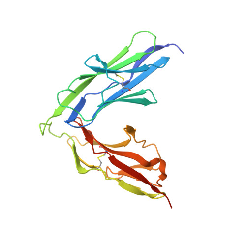
The Immunoglobulin-like Domains 1 and 2 of the Protein Tyrosine Phosphatase LAR Adopt an Unusual Horseshoe-like Conformation.

Biersmith, B.H.Hammel, M.Geisbrecht, E.R.Bouyain, S.

(2011) J Mol Biology 408: 616-627

  • DOI: https://doi.org/10.1016/j.jmb.2011.03.013
  • Primary Citation Related Structures: 
    3PXH, 3PXJ

  • PubMed Abstract: 

    Neurogenesis depends on exquisitely regulated interactions between macromolecules on the cell surface and in the extracellular matrix. In particular, interactions between proteoglycans and members of the type IIa subgroup of receptor protein tyrosine phosphatases underlie crucial developmental processes such as the formation of synapses at the neuromuscular junction and the migration of axons to their appropriate targets. We report the crystal structures of the first and second immunoglobulin-like domains of the Drosophila type IIa receptor Dlar and its mouse homolog LAR. These two domains adopt an unusual antiparallel arrangement that has not been reported in tandem repeats of immunoglobulin-like domains and that is presumably conserved in all type IIa receptor protein tyrosine phosphatases.


  • Organizational Affiliation
    • Division of Cell Biology and Biophysics, School of Biological Sciences, University of Missouri-Kansas City, 5100 Rockhill Road, Kansas City, MO 64110, USA.

Macromolecule Content 

  • Total Structure Weight: 90.97 kDa 
  • Atom Count: 6,103 
  • Modeled Residue Count: 779 
  • Deposited Residue Count: 840 
  • Unique protein chains: 1

Macromolecules

Find similar proteins by:|  3D Structure
Entity ID: 1
MoleculeChains  Sequence LengthOrganismDetailsImage
Tyrosine-protein phosphatase Lar
A, B, C, D
210Drosophila melanogasterMutation(s): 0 
Gene Names: LarCG10443
EC: 3.1.3.48
UniProt
Find proteins for P16621 (Drosophila melanogaster)
Explore P16621 
Go to UniProtKB:  P16621
Entity Groups
Sequence Clusters30% Identity50% Identity70% Identity90% Identity95% Identity100% Identity
UniProt GroupP16621
Sequence Annotations
Expand
Reference Sequence

Experimental Data & Validation

Experimental Data

  • Method: X-RAY DIFFRACTION
  • Resolution: 2.30 Å
  • R-Value Free:  0.261 (Depositor), 0.248 (DCC) 
  • R-Value Work:  0.223 (Depositor), 0.214 (DCC) 
  • R-Value Observed: 0.225 (Depositor) 
Space Group: P 1 21 1
Unit Cell:
Length ( Å )Angle ( ˚ )
a = 72.957α = 90
b = 77.513β = 101.08
c = 81.726γ = 90
Software Package:
Software NamePurpose
DENZOdata reduction
SCALEPACKdata scaling
PHASERphasing
PHENIXrefinement
PDB_EXTRACTdata extraction
SERGUIdata collection

Structure Validation

View Full Validation Report


Entry History 

Deposition Data

Revision History  (Full details and data files)

  • Version 1.0: 2011-03-23
    Type: Initial release
  • Version 1.1: 2011-07-13
    Changes: Version format compliance
  • Version 1.2: 2017-11-08
    Changes: Refinement description
  • Version 1.3: 2023-09-13
    Changes: Data collection, Database references, Refinement description
  • Version 1.4: 2024-10-30
    Changes: Structure summary