3PU8

PHF2 Jumonji-NOG-Fe(II) complex


Experimental Data Snapshot

  • Method: X-RAY DIFFRACTION
  • Resolution: 1.94 Å
  • R-Value Free: 0.235 
  • R-Value Work: 0.181 
  • R-Value Observed: 0.182 

wwPDB Validation   3D Report Full Report


This is version 1.3 of the entry. See complete history


Literature

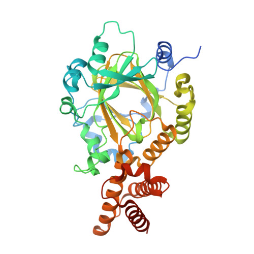
Structural basis for human PHF2 Jumonji domain interaction with metal ions.

Horton, J.R.Upadhyay, A.K.Hashimoto, H.Zhang, X.Cheng, X.

(2011) J Mol Biol 406: 1-8

  • DOI: https://doi.org/10.1016/j.jmb.2010.12.013
  • Primary Citation of Related Structures:  
    3PTR, 3PU3, 3PU8, 3PUA, 3PUS

  • PubMed Abstract: 

    PHF2 belongs to a class of α-ketoglutarate-Fe(2)(+)-dependent dioxygenases. PHF2 harbors a plant homeodomain (PHD) and a Jumonji domain. PHF2, via its PHD, binds Lys4-trimethylated histone 3 in submicromolar affinity and has been reported to have the demethylase activity of monomethylated lysine 9 of histone 3 in vivo. However, we did not detect demethylase activity for PHF2 Jumonji domain (with and without its linked PHD) in the context of histone peptides. We determined the crystal structures of PHF2 Jumonji domain in the absence and presence of additional exogenous metal ions. When Fe(2+) or Ni(2+) was added at a high concentration (50 mM) and allowed to soak in the preformed crystals, Fe(2+) or Ni(2+) was bound by six ligands in an octahedral coordination. The side chains of H249 and D251 and the two oxygen atoms of N-oxalylglycine (an analog of α-ketoglutarate) provide four coordinations in the equatorial plane, while the hydroxyl oxygen atom of Y321 and one water molecule provide the two axial coordinations as the fifth and sixth ligands, respectively. The metal binding site in PHF2 closely resembles the Fe(2+) sites in other Jumonji domains examined, with one important difference-a tyrosine (Y321 of PHF2) replaces histidine as the fifth ligand. However, neither Y321H mutation nor high metal concentration renders PHF2 an active demethylase on histone peptides. Wild type and Y321H mutant bind Ni(2+) with an approximately equal affinity of 50 μM. We propose that there must be other regulatory factors required for the enzymatic activity of PHF2 in vivo or that perhaps PHF2 acts on non-histone substrates. Furthermore, PHF2 shares significant sequence homology throughout the entire region, including the above-mentioned tyrosine at the corresponding iron-binding position, with that of Schizosaccharomyces pombe Epe1, which plays an essential role in heterochromatin function but has no known enzymatic activity.


  • Organizational Affiliation

    Department of Biochemistry, Emory University School of Medicine, Atlanta, GA 30322, USA.


Macromolecules
Find similar proteins by:  (by identity cutoff)  |  3D Structure
Entity ID: 1
MoleculeChains Sequence LengthOrganismDetailsImage
PHD finger protein 2A [auth B],
B [auth A]
392Homo sapiensMutation(s): 0 
Gene Names: PHF2KIAA0662
UniProt & NIH Common Fund Data Resources
Find proteins for O75151 (Homo sapiens)
Explore O75151 
Go to UniProtKB:  O75151
PHAROS:  O75151
GTEx:  ENSG00000197724 
Entity Groups  
Sequence Clusters30% Identity50% Identity70% Identity90% Identity95% Identity100% Identity
UniProt GroupO75151
Sequence Annotations
Expand
  • Reference Sequence
Small Molecules
Ligands 6 Unique
IDChains Name / Formula / InChI Key2D Diagram3D Interactions
OGA
Query on OGA

Download Ideal Coordinates CCD File 
BA [auth A],
J [auth B]
N-OXALYLGLYCINE
C4 H5 N O5
BIMZLRFONYSTPT-UHFFFAOYSA-N
SO4
Query on SO4

Download Ideal Coordinates CCD File 
EA [auth A],
FA [auth A],
M [auth B],
N [auth B]
SULFATE ION
O4 S
QAOWNCQODCNURD-UHFFFAOYSA-L
EDO
Query on EDO

Download Ideal Coordinates CCD File 
AA [auth A]
D [auth B]
E [auth B]
F [auth B]
G [auth B]
AA [auth A],
D [auth B],
E [auth B],
F [auth B],
G [auth B],
H [auth B],
I [auth B],
P [auth A],
Q [auth A],
R [auth A],
S [auth A],
T [auth A],
U [auth A],
V [auth A],
W [auth A],
X [auth A],
Y [auth A],
Z [auth A]
1,2-ETHANEDIOL
C2 H6 O2
LYCAIKOWRPUZTN-UHFFFAOYSA-N
FE
Query on FE

Download Ideal Coordinates CCD File 
DA [auth A],
L [auth B]
FE (III) ION
Fe
VTLYFUHAOXGGBS-UHFFFAOYSA-N
CL
Query on CL

Download Ideal Coordinates CCD File 
CA [auth A],
K [auth B]
CHLORIDE ION
Cl
VEXZGXHMUGYJMC-UHFFFAOYSA-M
OXY
Query on OXY

Download Ideal Coordinates CCD File 
C [auth B],
O [auth A]
OXYGEN MOLECULE
O2
MYMOFIZGZYHOMD-UHFFFAOYSA-N
Experimental Data & Validation

Experimental Data

  • Method: X-RAY DIFFRACTION
  • Resolution: 1.94 Å
  • R-Value Free: 0.235 
  • R-Value Work: 0.181 
  • R-Value Observed: 0.182 
  • Space Group: P 1 21 1
Unit Cell:
Length ( Å )Angle ( ˚ )
a = 65.748α = 90
b = 98.126β = 90.13
c = 66.517γ = 90
Software Package:
Software NamePurpose
HKL-2000data collection
PHASERphasing
PHENIXrefinement
HKL-2000data reduction
HKL-2000data scaling

Structure Validation

View Full Validation Report


Entry History 

Deposition Data

Revision History  (Full details and data files)

  • Version 1.0: 2011-01-26
    Type: Initial release
  • Version 1.1: 2011-07-13
    Changes: Version format compliance
  • Version 1.2: 2012-02-29
    Changes: Database references
  • Version 1.3: 2023-09-06
    Changes: Data collection, Database references, Derived calculations, Refinement description