


Multiple binding modes of a symmetric inhibitor in HIV-1 protease
Lindemann, I., Linde, K., Sammet, B., Heine, A., Diederich, W.E., Klebe, G.To be published.
Experimental Data Snapshot
Starting Model: experimental
View more details
Entity ID: 1 | |||||
|---|---|---|---|---|---|
| Molecule | Chains | Sequence Length | Organism | Details | Image |
| Protease | 99 | Human immunodeficiency virus type 1 (BRU ISOLATE) | Mutation(s): 0 EC: 3.4.23.16 | 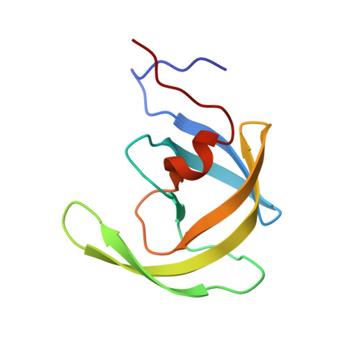 | |
UniProt | |||||
Entity Groups | |||||
| Sequence Clusters | 30% Identity50% Identity70% Identity90% Identity95% Identity100% Identity | ||||
| UniProt Group | P03366 | ||||
Sequence AnnotationsExpand | |||||
Reference Sequence | |||||
| Ligands 2 Unique | |||||
|---|---|---|---|---|---|
| ID | Chains | Name / Formula / InChI Key | 2D Diagram | 3D Interactions | |
| LJG Download:Ideal Coordinates CCD File | B [auth A] | N,N'-(iminodiethane-2,1-diyl)bis[4-amino-N-(2-methylpropyl)benzenesulfonamide] C24 H39 N5 O4 S2 BMXYVKCLBAGXOY-UHFFFAOYSA-N |  | ||
| CL Download:Ideal Coordinates CCD File | C [auth A] | CHLORIDE ION Cl VEXZGXHMUGYJMC-UHFFFAOYSA-M |  | ||
| Length ( Å ) | Angle ( ˚ ) |
|---|---|
| a = 62.652 | α = 90 |
| b = 62.652 | β = 90 |
| c = 82.438 | γ = 120 |
| Software Name | Purpose |
|---|---|
| MAR345dtb | data collection |
| PHASER | phasing |
| PHENIX | refinement |
| HKL-2000 | data reduction |
| HKL-2000 | data scaling |


