3PRZ | pdb_00003prz

Quinazolines with intra-molecular hydrogen bonding scaffold (iMHBS) as PI3K/mTOR dual inhibitors.


Experimental Data Snapshot

  • Method: X-RAY DIFFRACTION
  • Resolution: 2.60 Å
  • R-Value Free: 
    0.281 (Depositor), 0.280 (DCC) 
  • R-Value Work: 
    0.250 (Depositor), 0.240 (DCC) 
  • R-Value Observed: 
    0.251 (Depositor) 

wwPDB Validation   3D Report Full Report


Ligand Structure Quality Assessment 

Created with Raphaël 2.3.0Worse 01 BetterLigand structure goodness of fit to experimental dataBest fitted 3RZClick on this verticalbar to view details

This is version 1.2 of the entry. See complete history


Literature

Quinazolines with intra-molecular hydrogen bonding scaffold (iMHBS) as PI3K/mTOR dual inhibitors.

Liu, K.K.Huang, X.Bagrodia, S.Chen, J.H.Greasley, S.Cheng, H.Sun, S.Knighton, D.Rodgers, C.Rafidi, K.Zou, A.Xiao, J.Yan, S.

(2011) Bioorg Med Chem Lett 21: 1270-1274

  • DOI: https://doi.org/10.1016/j.bmcl.2010.12.026
  • Primary Citation of Related Structures:  
    3PRE, 3PRZ, 3PS6

  • PubMed Abstract: 

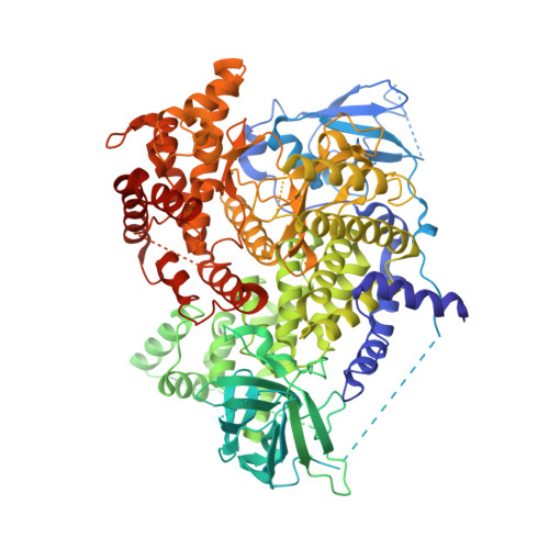
    Intra-molecular hydrogen bonding was introduced to the quinazoline motif to form a pseudo ring (intra-molecular H-bond scaffold, iMHBS) to mimic our previous published core structures, pyrido[2.3-D]pyrimidin-7-one and pteridinone, as PI3K/mTOR dual inhibitors. This design results in potent PI3K/mTOR dual inhibitors and the purposed intra-molecular hydrogen bonding structure is well supported by co-crystal structure in PI3Kγ enzyme. In addition, a novel synthetic route was developed for these analogs.


  • Organizational Affiliation

    Pfizer Global Research and Development, Chemistry Department, 10770 Science Center Drive, La Jolla, CA 92120, USA. Kevin.K.Liu@pfizer.com


Macromolecules
Find similar proteins by:  (by identity cutoff)  |  3D Structure
Entity ID: 1
MoleculeChains Sequence LengthOrganismDetailsImage
Phosphatidylinositol-4,5-bisphosphate 3-kinase catalytic subunit gamma isoform966Homo sapiensMutation(s): 0 
Gene Names: PIK3CG
EC: 2.7.1.153 (PDB Primary Data), 2.7.1.137 (UniProt), 2.7.1.154 (UniProt), 2.7.11.1 (UniProt)
UniProt & NIH Common Fund Data Resources
Find proteins for P48736 (Homo sapiens)
Explore P48736 
Go to UniProtKB:  P48736
PHAROS:  P48736
GTEx:  ENSG00000105851 
Entity Groups  
Sequence Clusters30% Identity50% Identity70% Identity90% Identity95% Identity100% Identity
UniProt GroupP48736
Sequence Annotations
Expand
  • Reference Sequence
Small Molecules
Ligands 1 Unique
IDChains Name / Formula / InChI Key2D Diagram3D Interactions
3RZ
Query on 3RZ

Download Ideal Coordinates CCD File 
B [auth A]4-amino-2-methyl-N-(1H-pyrazol-3-yl)quinazoline-8-carboxamide
C13 H12 N6 O
DMLHUAFVSLGSDJ-UHFFFAOYSA-N
Experimental Data & Validation

Experimental Data

  • Method: X-RAY DIFFRACTION
  • Resolution: 2.60 Å
  • R-Value Free:  0.281 (Depositor), 0.280 (DCC) 
  • R-Value Work:  0.250 (Depositor), 0.240 (DCC) 
  • R-Value Observed: 0.251 (Depositor) 
Space Group: C 1 2 1
Unit Cell:
Length ( Å )Angle ( ˚ )
a = 143.871α = 90
b = 67.188β = 95.65
c = 106.437γ = 90
Software Package:
Software NamePurpose
HKL-2000data collection
CNSrefinement
BUSTERrefinement
HKL-2000data reduction
SCALEPACKdata scaling
CNSphasing

Structure Validation

View Full Validation Report


Ligand Structure Quality Assessment 

Created with Raphaël 2.3.0Worse 01 BetterLigand structure goodness of fit to experimental dataBest fitted 3RZClick on this verticalbar to view details

Entry History 

Deposition Data

Revision History  (Full details and data files)

  • Version 1.0: 2011-02-09
    Type: Initial release
  • Version 1.1: 2011-07-13
    Changes: Version format compliance
  • Version 1.2: 2024-02-21
    Changes: Data collection, Database references, Derived calculations