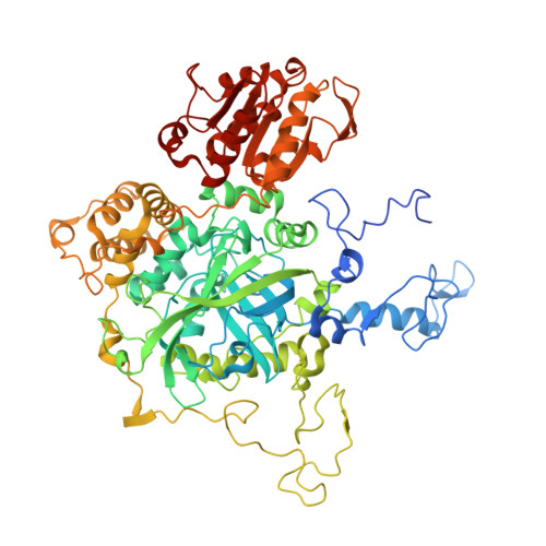
RCSB PDB - 3PQ7: Structure of I274C variant of E. coli KatE[] - Images 31-36

 3PQ7

Structure of I274C variant of E. coli KatE[] - Images 31-36


Experimental Data Snapshot

  • Method: X-RAY DIFFRACTION
  • Resolution: 1.80 Å
  • R-Value Free: 0.190 
  • R-Value Work: 0.142 
  • R-Value Observed: 0.144 

Starting Model: experimental
View more details

wwPDB Validation   3D Report Full Report


Ligand Structure Quality Assessment 

Created with Raphaël 2.3.0Worse 01 BetterLigand structure goodness of fit to experimental dataBest fitted HDEClick on this verticalbar to view detailsBest fitted HDDClick on this verticalbar to view details

This is version 1.5 of the entry. See complete history


Literature

Modulation of heme orientation and binding by a single residue in catalase HPII of Escherichia coli.

Jha, V.Louis, S.Chelikani, P.Carpena, X.Donald, L.J.Fita, I.Loewen, P.C.

(2011) Biochemistry 50: 2101-2110

  • DOI: https://doi.org/10.1021/bi200027v
  • Primary Citation of Related Structures:  
    3P9P, 3P9Q, 3P9R, 3P9S, 3PQ2, 3PQ3, 3PQ4, 3PQ5, 3PQ6, 3PQ7, 3PQ8

  • PubMed Abstract: 

    Heme-containing catalases have been extensively studied, revealing the roles of many residues, the existence of two heme orientations, flipped 180° relative to one another along the propionate-vinyl axis, and the presence of both heme b and heme d. The focus of this report is a residue, situated adjacent to the vinyl groups of the heme at the entrance of the lateral channel, with an unusual main chain geometry that is conserved in all catalase structures so far determined. In Escherichia coli catalase HPII, the residue is Ile274, and replacing it with Gly, Ala, and Val, found at the same location in other catalases, results in a reduction in catalytic efficiency, a reduced intensity of the Soret absorbance band, and a mixture of heme orientations and species. The reduced turnover rates and higher H(2)O(2) concentrations required to attain equivalent reaction velocities are explained in terms of less efficient containment of substrate H(2)O(2) in the heme cavity arising from easier escape through the more open entrance to the lateral channel created by the smaller side chains of Gly and Ala. Inserting a Cys at position 274 resulted in the heme being covalently linked to the protein through a Cys-vinyl bond that is hypersensitive to X-ray irradiation being largely degraded within seconds of exposure to the X-ray beam. Two heme orientations, flipped along the propionate-vinyl axis, are found in the Ala, Val, and Cys variants.


  • Organizational Affiliation

    Department of Microbiology, University of Manitoba, Winnipeg, MB R3T 2N2, Canada.


Macromolecules
Find similar proteins by:  (by identity cutoff)  |  3D Structure
Entity ID: 1
MoleculeChains Sequence LengthOrganismDetailsImage
Catalase HPII
A, B, C, D
753Escherichia coli K-12Mutation(s): 3 
Gene Names: b1732JW1721katE
EC: 1.11.1.6
UniProt
Find proteins for P21179 (Escherichia coli (strain K12))
Explore P21179 
Go to UniProtKB:  P21179
Entity Groups  
Sequence Clusters30% Identity50% Identity70% Identity90% Identity95% Identity100% Identity
UniProt GroupP21179
Sequence Annotations
Expand
  • Reference Sequence
Small Molecules
Ligands 3 Unique
IDChains Name / Formula / InChI Key2D Diagram3D Interactions
HDE
Query on HDE

Download Ideal Coordinates CCD File 
F [auth A],
I [auth B],
L [auth C],
O [auth D]
CIS-HEME D HYDROXYCHLORIN GAMMA-SPIROLACTONE 17R, 18S
C34 H38 Fe N4 O5
SQSYUADQFVTXKU-CAMYIWQSSA-N
HDD
Query on HDD

Download Ideal Coordinates CCD File 
E [auth A],
H [auth B],
K [auth C],
N [auth D]
CIS-HEME D HYDROXYCHLORIN GAMMA-SPIROLACTONE
C34 H32 Fe N4 O5
UMGOPAWIGKFTRK-QQDQPIDJSA-N
H2S
Query on H2S

Download Ideal Coordinates CCD File 
G [auth A],
J [auth B],
M [auth C],
P [auth D]
HYDROSULFURIC ACID
H2 S
RWSOTUBLDIXVET-UHFFFAOYSA-N
Experimental Data & Validation

Experimental Data

  • Method: X-RAY DIFFRACTION
  • Resolution: 1.80 Å
  • R-Value Free: 0.190 
  • R-Value Work: 0.142 
  • R-Value Observed: 0.144 
  • Space Group: P 1 21 1
Unit Cell:
Length ( Å )Angle ( ˚ )
a = 93.503α = 90
b = 133.022β = 109.4
c = 122.674γ = 90
Software Package:
Software NamePurpose
MOSFLMdata reduction
SCALAdata scaling
REFMACrefinement
PDB_EXTRACTdata extraction
MxDCdata collection
REFMACphasing

Structure Validation

View Full Validation Report


Ligand Structure Quality Assessment 

Created with Raphaël 2.3.0Worse 01 BetterLigand structure goodness of fit to experimental dataBest fitted HDEClick on this verticalbar to view detailsBest fitted HDDClick on this verticalbar to view details

Entry History 

Deposition Data

Revision History  (Full details and data files)

  • Version 1.0: 2010-12-22
    Type: Initial release
  • Version 1.1: 2011-07-13
    Changes: Version format compliance
  • Version 1.2: 2011-07-27
    Changes: Database references
  • Version 1.3: 2017-11-08
    Changes: Refinement description
  • Version 1.4: 2023-09-06
    Changes: Data collection, Database references, Derived calculations, Refinement description
  • Version 1.5: 2024-11-27
    Changes: Structure summary