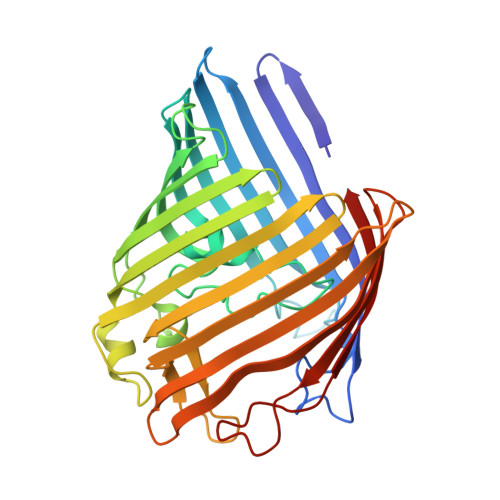
3POU

Crystal structure of E.coli OmpF porin in lipidic cubic phase: space group H32, large unit cell


Experimental Data Snapshot

  • Method: X-RAY DIFFRACTION
  • Resolution: 2.80 Å
  • R-Value Free: 0.252 
  • R-Value Work: 0.196 
  • R-Value Observed: 0.198 

Starting Model: experimental
View more details

wwPDB Validation   3D Report Full Report


Ligand Structure Quality Assessment 


This is version 1.2 of the entry. See complete history


Literature

Structure of Escherichia coli OmpF porin from lipidic mesophase.

Efremov, R.G.Sazanov, L.A.

(2012) J Struct Biol 178: 311-318

  • DOI: https://doi.org/10.1016/j.jsb.2012.03.005
  • Primary Citation of Related Structures:  
    3POQ, 3POU, 3POX

  • PubMed Abstract: 

    Outer membrane protein F, a major component of the Escherichia coli outer membrane, was crystallized for the first time in lipidic mesophase of monoolein in novel space groups, P1 and H32. Due to ease of its purification and crystallization OmpF can be used as a benchmark protein for establishing membrane protein crystallization in meso, as a "membrane lyzozyme". The packing of porin trimers in the crystals of space group H32 is similar to natural outer membranes, providing the first high-resolution insight into the close to native packing of OmpF. Surprisingly, interaction between trimers is mediated exclusively by lipids, without direct protein-protein contacts. Multiple ordered lipids are observed and many of them occupy identical positions independently of the space group, identifying preferential interaction sites of lipid acyl chains. Presence of ordered aliphatic chains close to a positively charged area on the porin surface suggests a position for a lipopolysaccharide binding site on the surface of the major E. coli porins.


  • Organizational Affiliation

    Medical Research Council Mitochondrial Biology Unit, Wellcome Trust/MRC Building, Hills Road, Cambridge CB2 0XY, UK.


Macromolecules
Find similar proteins by:  (by identity cutoff)  |  3D Structure
Entity ID: 1
MoleculeChains Sequence LengthOrganismDetailsImage
OmpF protein340Escherichia coli BL21Mutation(s): 0 
Membrane Entity: Yes 
UniProt
Find proteins for P02931 (Escherichia coli (strain K12))
Explore P02931 
Go to UniProtKB:  P02931
Entity Groups  
Sequence Clusters30% Identity50% Identity70% Identity90% Identity95% Identity100% Identity
UniProt GroupP02931
Sequence Annotations
Expand
  • Reference Sequence
Experimental Data & Validation

Experimental Data

  • Method: X-RAY DIFFRACTION
  • Resolution: 2.80 Å
  • R-Value Free: 0.252 
  • R-Value Work: 0.196 
  • R-Value Observed: 0.198 
  • Space Group: H 3 2
Unit Cell:
Length ( Å )Angle ( ˚ )
a = 85.71α = 90
b = 85.71β = 90
c = 332.941γ = 120
Software Package:
Software NamePurpose
MOSFLMdata reduction
SCALAdata scaling
PHASERphasing
REFMACrefinement
PDB_EXTRACTdata extraction

Structure Validation

View Full Validation Report


Ligand Structure Quality Assessment 


Entry History 

Deposition Data

Revision History  (Full details and data files)

  • Version 1.0: 2012-03-07
    Type: Initial release
  • Version 1.1: 2012-06-20
    Changes: Database references
  • Version 1.2: 2023-11-01
    Changes: Data collection, Database references, Derived calculations, Refinement description