3POC | pdb_00003poc

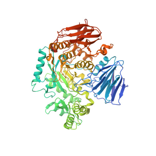
The crystal structure of the D307A mutant of alpha-Glucosidase (FAMILY 31) from Ruminococcus obeum ATCC 29174 in complex with acarbose


Experimental Data Snapshot

  • Method: X-RAY DIFFRACTION
  • Resolution: 1.99 Å
  • R-Value Free: 
    0.225 (Depositor), 0.206 (DCC) 
  • R-Value Work: 
    0.168 (Depositor) 
  • R-Value Observed: 
    0.171 (Depositor) 

Starting Model: experimental
View more details

wwPDB Validation 3D Report Full Report

Validation slider image for 3POC

Ligand Structure Quality Assessment 


This is version 2.2 of the entry. See complete history. 

Literature

Interaction of antidiabetic alpha-glucosidase inhibitors and gut bacteria alpha-glucosidase.

Tan, K., Tesar, C., Wilton, R., Jedrzejczak, R.P., Joachimiak, A.

(2018) Protein Sci 27: 1498-1508

  • DOI: https://doi.org/10.1002/pro.3444
  • Primary Citation Related Structures: 
    3PHA, 3POC, 6C9X, 6C9Z, 6CA1, 6CA3

  • PubMed Abstract: 

    Carbohydrate hydrolyzing α-glucosidases are commonly found in microorganisms present in the human intestine microbiome. We have previously reported crystal structures of an α-glucosidase from the human gut bacterium Blaubia (Ruminococcus) obeum (Ro-αG1) and its substrate preference/specificity switch. This novel member of the GH31 family is a structural homolog of human intestinal maltase-glucoamylase (MGAM) and sucrase-isomaltase (SI) with a highly conserved active site that is predicted to be common in Ro-αG1 homologs among other species that colonize the human gut. In this report, we present structures of Ro-αG1 in complex with the antidiabetic α-glucosidase inhibitors voglibose, miglitol, and acarbose and supporting binding data. The in vitro binding of these antidiabetic drugs to Ro-αG1 suggests the potential for unintended in vivo crossreaction of the α-glucosidase inhibitors to bacterial α-glucosidases that are present in gut microorganism communities. Moreover, analysis of these drug-bound enzyme structures could benefit further antidiabetic drug development.


  • Organizational Affiliation: 
    • Midwest Center for Structural Genomics and Structural Biology Center, Biosciences, Argonne National Laboratory, Argonne, Illinois, 60439.

Macromolecule Content 

  • Total Structure Weight: 156.37 kDa 
  • Atom Count: 11,497 
  • Modeled Residue Count: 1,328 
  • Deposited Residue Count: 1,332 
  • Unique protein chains: 1

Macromolecules

Find similar proteins by:|  3D Structure
Entity ID: 1
MoleculeChains  Sequence LengthOrganismDetailsImage
alpha-Glucosidase
A, B
666Blautia obeum ATCC 29174Mutation(s): 1 
Gene Names: RUMOBE_03919
EC: 3.2.1.3
UniProt
Find proteins for A5ZY13 (Blautia obeum ATCC 29174)
Explore A5ZY13 
Go to UniProtKB:  A5ZY13
Entity Groups
Sequence Clusters30% Identity50% Identity70% Identity90% Identity95% Identity100% Identity
UniProt GroupA5ZY13
Sequence Annotations
Expand
Reference Sequence

Oligosaccharides

Help  
Entity ID: 2
MoleculeChains Length2D Diagram GlycosylationD Interactions
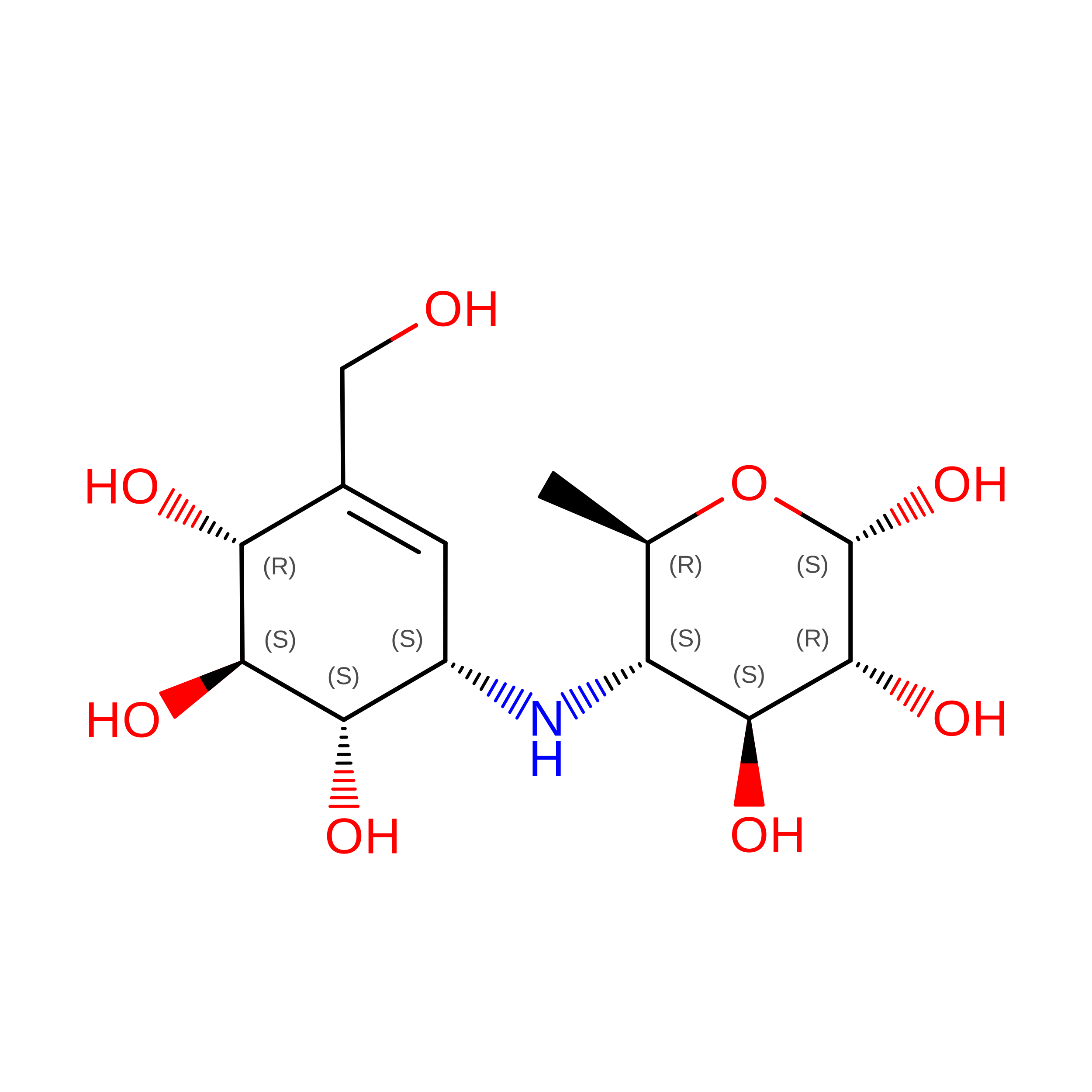
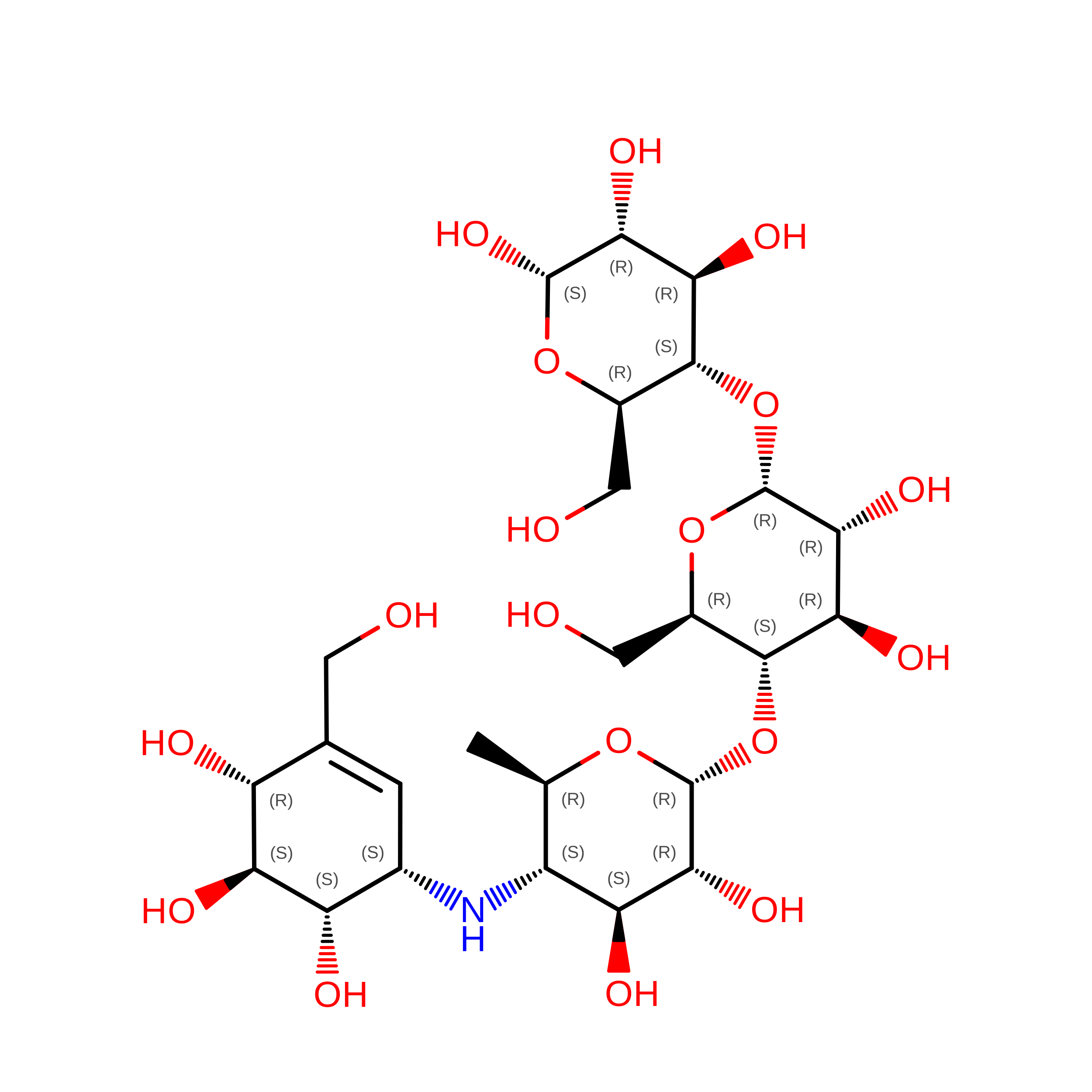
4,6-dideoxy-4-{[(1S,4R,5S,6S)-4,5,6-trihydroxy-3-(hydroxymethyl)cyclohex-2-en-1-yl]amino}-alpha-D-glucopyranose-(1-4)-alpha-D-glucopyranose-(1-4)-alpha-D-glucopyranose
C
3N/AN/A
Glycosylation Resources
GlyTouCan: G66431MI
GlyCosmos: G66431MI

Biologically Interesting Molecules (External Reference) 

1 Unique

Experimental Data & Validation

Experimental Data

  • Method: X-RAY DIFFRACTION
  • Resolution: 1.99 Å
  • R-Value Free:  0.225 (Depositor), 0.206 (DCC) 
  • R-Value Work:  0.168 (Depositor) 
  • R-Value Observed: 0.171 (Depositor) 
Space Group: P 1 21 1
Unit Cell:
Length ( Å )Angle ( ˚ )
a = 67.954α = 90
b = 125.691β = 107.62
c = 87.968γ = 90
Software Package:
Software NamePurpose
SBC-Collectdata collection
MOLREPphasing
PHENIXrefinement
HKL-3000data reduction
HKL-3000data scaling

Structure Validation

View Full Validation Report


Ligand Structure Quality Assessment 


Entry History 

Deposition Data

Revision History  (Full details and data files)

  • Version 1.0: 2011-01-26
    Type: Initial release
  • Version 1.1: 2011-07-13
    Changes: Version format compliance
  • Version 2.0: 2020-07-29
    Type: Remediation
    Reason: Carbohydrate remediation
    Changes: Advisory, Atomic model, Data collection, Database references, Derived calculations, Non-polymer description, Structure summary
  • Version 2.1: 2023-09-06
    Changes: Data collection, Database references, Refinement description, Structure summary
  • Version 2.2: 2026-09-30
    Changes: Database references, Structure summary