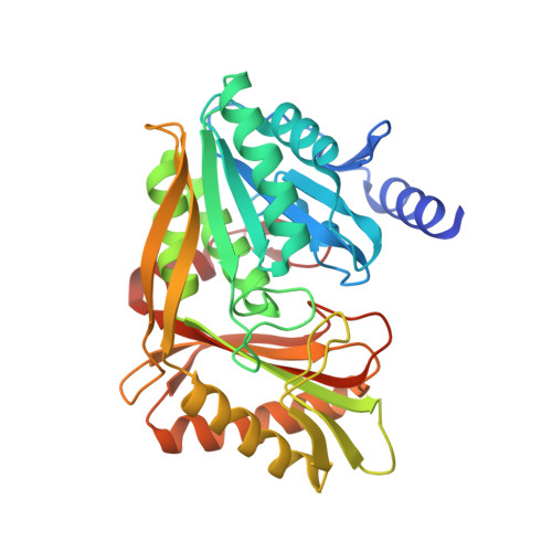
3PNQ

Crystal Structure of E.coli Dha kinase DhaK (H56N) complex with Dha


Experimental Data Snapshot

  • Method: X-RAY DIFFRACTION
  • Resolution: 2.20 Å
  • R-Value Free: 0.220 
  • R-Value Work: 0.200 
  • R-Value Observed: 0.201 

wwPDB Validation   3D Report Full Report


This is version 1.3 of the entry. See complete history


Literature

Structural and mechanistic insight into covalent substrate binding by Escherichia coli dihydroxyacetone kinase.

Shi, R.McDonald, L.Cui, Q.Matte, A.Cygler, M.Ekiel, I.

(2011) Proc Natl Acad Sci U S A 108: 1302-1307

  • DOI: https://doi.org/10.1073/pnas.1012596108
  • Primary Citation of Related Structures:  
    3PNK, 3PNL, 3PNM, 3PNO, 3PNQ

  • PubMed Abstract: 

    The Escherichia coli dihydroxyacetone (Dha) kinase is an unusual kinase because (i) it uses the phosphoenolpyruvate carbohydrate: phosphotransferase system (PTS) as the source of high-energy phosphate, (ii) the active site is formed by two subunits, and (iii) the substrate is covalently bound to His218(K)* of the DhaK subunit. The PTS transfers phosphate to DhaM, which in turn phosphorylates the permanently bound ADP coenzyme of DhaL. This phosphoryl group is subsequently transferred to the Dha substrate bound to DhaK. Here we report the crystal structure of the E. coli Dha kinase complex, DhaK-DhaL. The structure of the complex reveals that DhaK undergoes significant conformational changes to accommodate binding of DhaL. Combined mutagenesis and enzymatic activity studies of kinase mutants allow us to propose a catalytic mechanism for covalent Dha binding, phosphorylation, and release of the Dha-phosphate product. Our results show that His56(K) is involved in formation of the covalent hemiaminal bond with Dha. The structure of H56N(K) with noncovalently bound substrate reveals a somewhat different positioning of Dha in the binding pocket as compared to covalently bound Dha, showing that the covalent attachment to His218(K) orients the substrate optimally for phosphoryl transfer. Asp109(K) is critical for activity, likely acting as a general base activating the γ-OH of Dha. Our results provide a comprehensive picture of the roles of the highly conserved active site residues of dihydroxyacetone kinases.


  • Organizational Affiliation

    Department of Biochemistry, McGill University, Montréal, QC, Canada H3G 1Y6.


Macromolecules
Find similar proteins by:  (by identity cutoff)  |  3D Structure
Entity ID: 1
MoleculeChains Sequence LengthOrganismDetailsImage
PTS-dependent dihydroxyacetone kinase, dihydroxyacetone-binding subunit dhaK
A, B, C, D
357Escherichia coli K-12Mutation(s): 1 
Gene Names: b1200dhaKJW5187ycgT
EC: 2.7 (PDB Primary Data), 2.7.1.121 (UniProt)
UniProt
Find proteins for P76015 (Escherichia coli (strain K12))
Explore P76015 
Go to UniProtKB:  P76015
Entity Groups  
Sequence Clusters30% Identity50% Identity70% Identity90% Identity95% Identity100% Identity
UniProt GroupP76015
Sequence Annotations
Expand
  • Reference Sequence
Small Molecules
Ligands 1 Unique
IDChains Name / Formula / InChI Key2D Diagram3D Interactions
2HA
Query on 2HA

Download Ideal Coordinates CCD File 
E [auth D]Dihydroxyacetone
C3 H6 O3
RXKJFZQQPQGTFL-UHFFFAOYSA-N
Experimental Data & Validation

Experimental Data

  • Method: X-RAY DIFFRACTION
  • Resolution: 2.20 Å
  • R-Value Free: 0.220 
  • R-Value Work: 0.200 
  • R-Value Observed: 0.201 
  • Space Group: P 1 21 1
Unit Cell:
Length ( Å )Angle ( ˚ )
a = 82.232α = 90
b = 101.073β = 89.95
c = 99.349γ = 90
Software Package:
Software NamePurpose
DENZOdata reduction
SCALEPACKdata scaling
REFMACrefinement
PDB_EXTRACTdata extraction
HKL-2000data scaling
MOLREPphasing

Structure Validation

View Full Validation Report


Entry History 

Deposition Data

Revision History  (Full details and data files)

  • Version 1.0: 2011-01-12
    Type: Initial release
  • Version 1.1: 2011-07-13
    Changes: Version format compliance
  • Version 1.2: 2017-11-08
    Changes: Refinement description
  • Version 1.3: 2024-02-21
    Changes: Data collection, Database references, Derived calculations, Structure summary