3PGM | pdb_00003pgm

THE STRUCTURE OF YEAST PHOSPHOGLYCERATE MUTASE AT 0.28 NM RESOLUTION


Experimental Data Snapshot

  • Method: X-RAY DIFFRACTION
  • Resolution: 2.80 Å

wwPDB Validation 3D Report Full Report

Validation slider image for 3PGM

This is version 1.6 of the entry. See complete history

Literature

Structure and activity of phosphoglycerate mutase.

Winn, S.I.Watson, H.C.Harkins, R.N.Fothergill, L.A.

(1981) Philos Trans R Soc London,ser B 293: 121-130

  • DOI: https://doi.org/10.1098/rstb.1981.0066
  • Primary Citation Related Structures: 
    3PGM

  • PubMed Abstract: 

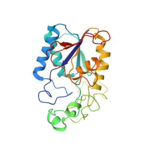
    The structure of yeast phosphoglycerate mutase determined by X-ray crystallographic and amino acid sequence studies has been interpreted in terms of the chemical, kinetic and mechanistic observations made on this enzyme. There are two histidine residues at the active site, with imidazole groups almost parallel to each other and approximately 0.4 nm apart, positioned close to the 2 and 3 positions of the substrate. The simplest interpretation of the available information suggests that a ping-pong type mechanism operates in which at least one of these histidine residues participates in the phosphoryl transfer reaction. The flexible C-terminal region also plays an important role in the enzymic reaction.

Macromolecule Content 

  • Total Structure Weight: 55.09 kDa 
  • Atom Count: 3,708 
  • Modeled Residue Count: 460 
  • Deposited Residue Count: 488 
  • Unique protein chains: 1

Macromolecules

Find similar proteins by:|  3D Structure
Entity ID: 1
MoleculeChains  Sequence LengthOrganismDetailsImage
Phosphoglycerate mutase 1
A, B
244Saccharomyces cerevisiaeMutation(s): 0 
EC: 2.7.5.3 (PDB Primary Data), 5.4.2.11 (UniProt)
UniProt
Find proteins for P00950 (Saccharomyces cerevisiae (strain ATCC 204508 / S288c))
Explore P00950 
Go to UniProtKB:  P00950
Entity Groups
Sequence Clusters30% Identity50% Identity70% Identity90% Identity95% Identity100% Identity
UniProt GroupP00950
Sequence Annotations
Expand
Reference Sequence

Experimental Data & Validation

Experimental Data

  • Method: X-RAY DIFFRACTION
  • Resolution: 2.80 Å
Space Group: C 1 2 1
Unit Cell:
Length ( Å )Angle ( ˚ )
a = 96.4α = 90
b = 85.9β = 120.6
c = 81.9γ = 90

Structure Validation

View Full Validation Report


Entry History 

Deposition Data

Revision History  (Full details and data files)

  • Version 1.0: 1982-05-26
    Type: Initial release
  • Version 1.1: 2008-03-03
    Changes: Version format compliance
  • Version 1.2: 2011-07-13
    Changes: Version format compliance
  • Version 1.3: 2013-08-14
    Changes: Other
  • Version 1.4: 2017-11-29
    Changes: Derived calculations, Other
  • Version 1.5: 2023-05-31
    Changes: Database references, Derived calculations
  • Version 1.6: 2024-05-22
    Changes: Data collection