3PG0 | pdb_00003pg0

Crystal structure of designed 3-fold symmetric protein, ThreeFoil


Experimental Data Snapshot

  • Method: X-RAY DIFFRACTION
  • Resolution: 1.62 Å
  • R-Value Free: 
    0.184 (Depositor), 0.177 (DCC) 
  • R-Value Work: 
    0.166 (Depositor), 0.159 (DCC) 
  • R-Value Observed: 
    0.167 (Depositor) 

Starting Model: experimental
View more details

wwPDB Validation 3D Report Full Report

Validation slider image for 3PG0

This is version 1.3 of the entry. See complete history

Literature

Modular evolution and the origins of symmetry: reconstruction of a three-fold symmetric globular protein.

Broom, A.Doxey, A.C.Lobsanov, Y.D.Berthin, L.G.Rose, D.R.Howell, P.L.McConkey, B.J.Meiering, E.M.

(2012) Structure 20: 161-171

  • DOI: https://doi.org/10.1016/j.str.2011.10.021
  • Primary Citation Related Structures: 
    3PG0

  • PubMed Abstract: 

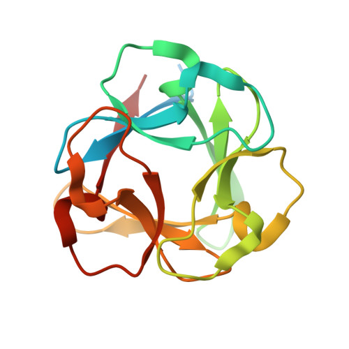
    The high frequency of internal structural symmetry in common protein folds is presumed to reflect their evolutionary origins from the repetition and fusion of ancient peptide modules, but little is known about the primary sequence and physical determinants of this process. Unexpectedly, a sequence and structural analysis of symmetric subdomain modules within an abundant and ancient globular fold, the β-trefoil, reveals that modular evolution is not simply a relic of the ancient past, but is an ongoing and recurring mechanism for regenerating symmetry, having occurred independently in numerous existing β-trefoil proteins. We performed a computational reconstruction of a β-trefoil subdomain module and repeated it to form a newly three-fold symmetric globular protein, ThreeFoil. In addition to its near perfect structural identity between symmetric modules, ThreeFoil is highly soluble, performs multivalent carbohydrate binding, and has remarkably high thermal stability. These findings have far-reaching implications for understanding the evolution and design of proteins via subdomain modules.


  • Organizational Affiliation
    • Guelph-Waterloo Centre for Graduate Studies in Chemistry and Biochemistry, University of Waterloo, 200 University Avenue West, Waterloo, Ontario, N2L 3G1, Canada.

Macromolecule Content 

  • Total Structure Weight: 18.9 kDa 
  • Atom Count: 1,327 
  • Modeled Residue Count: 140 
  • Deposited Residue Count: 165 
  • Unique protein chains: 1

Macromolecules

Find similar proteins by:|  3D Structure
Entity ID: 1
MoleculeChains  Sequence LengthOrganismDetailsImage
ThreeFoil165synthetic constructMutation(s): 0 

Experimental Data & Validation

Experimental Data

  • Method: X-RAY DIFFRACTION
  • Resolution: 1.62 Å
  • R-Value Free:  0.184 (Depositor), 0.177 (DCC) 
  • R-Value Work:  0.166 (Depositor), 0.159 (DCC) 
  • R-Value Observed: 0.167 (Depositor) 
Space Group: P 43 21 2
Unit Cell:
Length ( Å )Angle ( ˚ )
a = 45.012α = 90
b = 45.012β = 90
c = 113.439γ = 90
Software Package:
Software NamePurpose
d*TREKdata scaling
PHENIXrefinement
PDB_EXTRACTdata extraction
CrystalCleardata collection
d*TREKdata reduction
BALBESphasing
MOLREPphasing

Structure Validation

View Full Validation Report


Entry History 

Deposition Data

Revision History  (Full details and data files)

  • Version 1.0: 2011-12-21
    Type: Initial release
  • Version 1.1: 2012-02-01
    Changes: Database references
  • Version 1.2: 2017-11-08
    Changes: Refinement description
  • Version 1.3: 2023-09-06
    Changes: Data collection, Database references, Derived calculations, Refinement description