3PCN | pdb_00003pcn

STRUCTURE OF PROTOCATECHUATE 3,4-DIOXYGENASE COMPLEXED WITH 3,4-DIHYDROXYPHENYLACETATE


Experimental Data Snapshot

  • Method: X-RAY DIFFRACTION
  • Resolution: 2.40 Å
  • R-Value Work: 
    0.166 (Depositor) 

wwPDB Validation 3D Report Full Report

Validation slider image for 3PCN

This is version 2.0 of the entry. See complete history

Literature

Crystal structure and resonance Raman studies of protocatechuate 3,4-dioxygenase complexed with 3,4-dihydroxyphenylacetate.

Elgren, T.E.Orville, A.M.Kelly, K.A.Lipscomb, J.D.Ohlendorf, D.H.Que Jr., L.

(1997) Biochemistry 36: 11504-11513

  • DOI: https://doi.org/10.1021/bi970691k
  • Primary Citation Related Structures: 
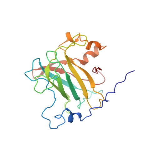
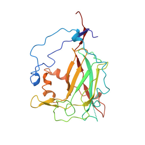
    3PCN

  • PubMed Abstract: 

    The crystal structure of the anaerobic complex of Pseudomonas putida protocatechuate 3,4-dioxygenase (3,4-PCD) bound with the alternative substrate, 3,4-dihydroxyphenylacetate (HPCA), is reported at 2.4 A resolution and refined to an R factor of 0.17. Formation of the active site Fe(III).HPCA chelated complex causes the endogenous axial tyrosinate, Tyr447 (147beta), to dissociate from the iron and rotate into an alternative orientation analogous to that previously observed in the anaerobic 3,4-PCD.3,4-dihydroxybenzoate complex (3, 4-PCD.PCA) [Orville, A. M., Lipscomb, J. D., & Ohlendorf, D. H. (1997) Biochemistry 36, 10052-10066]. Two orientations of the aromatic ring of HPCA related by an approximate 180 degrees rotation within the active site are consistent with the electron density. Resonance Raman (rR) spectroscopic data from Brevibacteriumfuscum 3,4-PCD.HPCA complex in solution reveals low frequency rR vibrational bands between 500 and 650 cm-1 as well as a band at approximately 1320 cm-1 which are diagnostic of a HPCA. Fe(III) chelate complex. 18O labeling of HPCA at either the C4 or C3 hydroxyl group unambiguously establishes the vibrational coupling modes associated with the five-membered chelate ring system. Analysis of these data suggests that the Fe(III)-HPCAO4 bond is shorter than the Fe(III)-HPCAO3 bond. This consequently favors the model for the crystal structure in which the C3 phenolic function occupies the Fe3+ ligand site opposite the endogenous ligand Tyr408(Oeta) (108beta). This is essentially the same binding orientation as proposed for PCA in the crystal structure of the anaerobic 3,4-PCD.PCA complex based solely on direct modeling of the 2Fo - Fc electron density and suggests that this is the conformation required for catalysis.


  • Organizational Affiliation
    • Department of Chemistry, Hamilton College, Clinton, New York 13323, USA.

Macromolecule Content 

  • Total Structure Weight: 295.66 kDa 
  • Atom Count: 22,014 
  • Modeled Residue Count: 2,598 
  • Deposited Residue Count: 2,628 
  • Unique protein chains: 2

Macromolecules

Find similar proteins by:|  3D Structure
Entity ID: 1
MoleculeChains  Sequence LengthOrganismDetailsImage
PROTOCATECHUATE 3,4-DIOXYGENASE200Pseudomonas putidaMutation(s): 0 
EC: 1.13.11.3
UniProt
Find proteins for P00436 (Pseudomonas putida)
Explore P00436 
Go to UniProtKB:  P00436
Entity Groups
Sequence Clusters30% Identity50% Identity70% Identity90% Identity95% Identity100% Identity
UniProt GroupP00436
Sequence Annotations
Expand
Reference Sequence
Find similar proteins by:|  3D Structure
Entity ID: 2
MoleculeChains  Sequence LengthOrganismDetailsImage
PROTOCATECHUATE 3,4-DIOXYGENASE238Pseudomonas putidaMutation(s): 0 
EC: 1.13.11.3
UniProt
Find proteins for P00437 (Pseudomonas putida)
Explore P00437 
Go to UniProtKB:  P00437
Entity Groups
Sequence Clusters30% Identity50% Identity70% Identity90% Identity95% Identity100% Identity
UniProt GroupP00437
Sequence Annotations
Expand
Reference Sequence

Small Molecules

Ligands 3 Unique
IDChains Name / Formula / InChI Key2D Diagram3D Interactions
DHY

Query on DHY


Download:Ideal Coordinates CCD File
AA [auth Q]
DA [auth R]
O [auth M]
R [auth N]
U [auth O]
AA [auth Q],
DA [auth R],
O [auth M],
R [auth N],
U [auth O],
X [auth P]
2-(3,4-DIHYDROXYPHENYL)ACETIC ACID
C8 H8 O4
CFFZDZCDUFSOFZ-UHFFFAOYSA-N
BME

Query on BME


Download:Ideal Coordinates CCD File
CA [auth R]
N [auth M]
Q [auth N]
T [auth O]
W [auth P]
CA [auth R],
N [auth M],
Q [auth N],
T [auth O],
W [auth P],
Z [auth Q]
BETA-MERCAPTOETHANOL
C2 H6 O S
DGVVWUTYPXICAM-UHFFFAOYSA-N
FE

Query on FE


Download:Ideal Coordinates CCD File
BA [auth R]
M
P [auth N]
S [auth O]
V [auth P]
BA [auth R],
M,
P [auth N],
S [auth O],
V [auth P],
Y [auth Q]
FE (III) ION
Fe
VTLYFUHAOXGGBS-UHFFFAOYSA-N

Experimental Data & Validation

Experimental Data

  • Method: X-RAY DIFFRACTION
  • Resolution: 2.40 Å
  • R-Value Work:  0.166 (Depositor) 
Space Group: I 1 2 1
Unit Cell:
Length ( Å )Angle ( ˚ )
a = 196.6α = 90
b = 127.5β = 97.7
c = 134.3γ = 90
Software Package:
Software NamePurpose
PROLSQrefinement
XENGENdata reduction
XENGENdata scaling

Structure Validation

View Full Validation Report


Entry History 

Deposition Data

Revision History  (Full details and data files)

  • Version 1.0: 1998-02-25
    Type: Initial release
  • Version 1.1: 2008-03-03
    Changes: Version format compliance
  • Version 1.2: 2011-07-13
    Changes: Advisory, Derived calculations, Version format compliance
  • Version 2.0: 2023-09-27
    Type: Remediation
    Changes: Advisory, Atomic model, Data collection, Database references, Derived calculations, Other, Refinement description