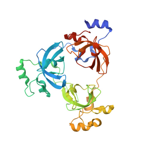
3P8H

Crystal structure of L3MBTL1 (MBT repeat) in complex with a nicotinamide antagonist


Experimental Data Snapshot

  • Method: X-RAY DIFFRACTION
  • Resolution: 2.55 Å
  • R-Value Free: 0.242 
  • R-Value Work: 0.191 
  • R-Value Observed: 0.194 

Starting Model: experimental
View more details

wwPDB Validation   3D Report Full Report


Ligand Structure Quality Assessment 


This is version 1.3 of the entry. See complete history


Literature

Small-molecule ligands of methyl-lysine binding proteins.

Herold, J.M.Wigle, T.J.Norris, J.L.Lam, R.Korboukh, V.K.Gao, C.Ingerman, L.A.Kireev, D.B.Senisterra, G.Vedadi, M.Tripathy, A.Brown, P.J.Arrowsmith, C.H.Jin, J.Janzen, W.P.Frye, S.V.

(2011) J Med Chem 54: 2504-2511

  • DOI: https://doi.org/10.1021/jm200045v
  • Primary Citation of Related Structures:  
    3P8H

  • PubMed Abstract: 

    Proteins which bind methylated lysines ("readers" of the histone code) are important components in the epigenetic regulation of gene expression and can also modulate other proteins that contain methyl-lysine such as p53 and Rb. Recognition of methyl-lysine marks by MBT domains leads to compaction of chromatin and a repressed transcriptional state. Antagonists of MBT domains would serve as probes to interrogate the functional role of these proteins and initiate the chemical biology of methyl-lysine readers as a target class. Small-molecule MBT antagonists were designed based on the structure of histone peptide-MBT complexes and their interaction with MBT domains determined using a chemiluminescent assay and ITC. The ligands discovered antagonize native histone peptide binding, exhibiting 5-fold stronger binding affinity to L3MBTL1 than its preferred histone peptide. The first cocrystal structure of a small molecule bound to L3MBTL1 was determined and provides new insights into binding requirements for further ligand design.


  • Organizational Affiliation

    Center for Integrated Chemical Biology and Drug Discovery, UNC Eshelman School of Pharmacy, Division of Medicinal Chemistry and Natural Products, University of North Carolina at Chapel Hill, Chapel Hill, North Carolina 27599, United States.


Macromolecules
Find similar proteins by:  (by identity cutoff)  |  3D Structure
Entity ID: 1
MoleculeChains Sequence LengthOrganismDetailsImage
Lethal(3)malignant brain tumor-like protein
A, B, C
323Homo sapiensMutation(s): 0 
Gene Names: KIAA0681L3MBTL3MBTLL3MBTL1
UniProt & NIH Common Fund Data Resources
Find proteins for Q9Y468 (Homo sapiens)
Explore Q9Y468 
Go to UniProtKB:  Q9Y468
PHAROS:  Q9Y468
GTEx:  ENSG00000185513 
Entity Groups  
Sequence Clusters30% Identity50% Identity70% Identity90% Identity95% Identity100% Identity
UniProt GroupQ9Y468
Sequence Annotations
Expand
  • Reference Sequence
Small Molecules
Ligands 3 Unique
IDChains Name / Formula / InChI Key2D Diagram3D Interactions
P8H
Query on P8H

Download Ideal Coordinates CCD File 
D [auth A],
H [auth B]
3-bromo-5-[(4-pyrrolidin-1-ylpiperidin-1-yl)carbonyl]pyridine
C15 H20 Br N3 O
CQERVFFAOOUFEQ-UHFFFAOYSA-N
SO4
Query on SO4

Download Ideal Coordinates CCD File 
E [auth A],
I [auth B],
J [auth C]
SULFATE ION
O4 S
QAOWNCQODCNURD-UHFFFAOYSA-L
GOL
Query on GOL

Download Ideal Coordinates CCD File 
F [auth A],
G [auth A]
GLYCEROL
C3 H8 O3
PEDCQBHIVMGVHV-UHFFFAOYSA-N
Binding Affinity Annotations 
IDSourceBinding Affinity
P8H BindingDB:  3P8H Kd: min: 5000, max: 1.00e+4 (nM) from 2 assay(s)
IC50: min: 4200, max: 2.10e+4 (nM) from 3 assay(s)
PDBBind:  3P8H Kd: 5000 (nM) from 1 assay(s)
Experimental Data & Validation

Experimental Data

  • Method: X-RAY DIFFRACTION
  • Resolution: 2.55 Å
  • R-Value Free: 0.242 
  • R-Value Work: 0.191 
  • R-Value Observed: 0.194 
  • Space Group: P 32
Unit Cell:
Length ( Å )Angle ( ˚ )
a = 106.264α = 90
b = 106.264β = 90
c = 90.144γ = 120
Software Package:
Software NamePurpose
DENZOdata reduction
SCALEPACKdata scaling
MOLREPphasing
REFMACrefinement
PDB_EXTRACTdata extraction
MxDCdata collection
HKL-2000data reduction
HKL-2000data scaling

Structure Validation

View Full Validation Report


Ligand Structure Quality Assessment 


Entry History 

Revision History  (Full details and data files)

  • Version 1.0: 2010-11-03
    Type: Initial release
  • Version 1.1: 2011-07-13
    Changes: Version format compliance
  • Version 1.2: 2017-11-08
    Changes: Refinement description
  • Version 1.3: 2023-09-06
    Changes: Data collection, Database references, Derived calculations, Refinement description