3P5N | pdb_00003p5n

Structure and mechanism of the S component of a bacterial ECF transporter


Experimental Data Snapshot

  • Method: X-RAY DIFFRACTION
  • Resolution: 3.60 Å
  • R-Value Free: 
    0.289 (Depositor), 0.290 (DCC) 
  • R-Value Work: 
    0.265 (Depositor), 0.270 (DCC) 
  • R-Value Observed: 
    0.266 (Depositor) 

wwPDB Validation 3D Report Full Report

Validation slider image for 3P5N

Ligand Structure Quality Assessment 


This is version 1.3 of the entry. See complete history

Literature

Structure and mechanism of the S component of a bacterial ECF transporter

Zhang, P.Wang, J.Shi, Y.

(2010) Nature 468: 717-720

  • DOI: https://doi.org/10.1038/nature09488
  • Primary Citation Related Structures: 
    3P5N

  • PubMed Abstract: 

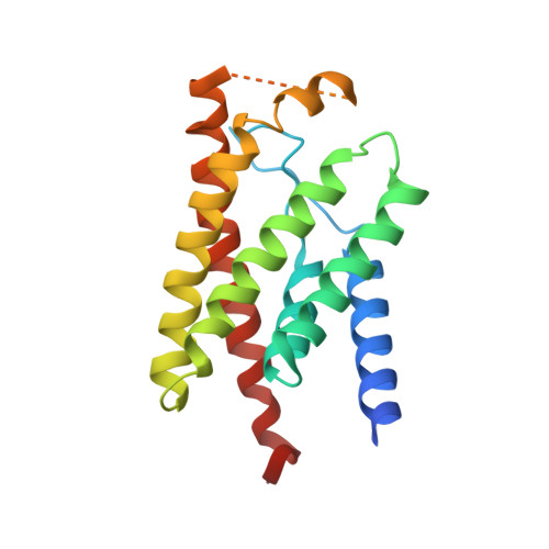
    The energy-coupling factor (ECF) transporters, responsible for vitamin uptake in prokaryotes, are a unique family of membrane transporters. Each ECF transporter contains a membrane-embedded, substrate-binding protein (known as the S component), an energy-coupling module that comprises two ATP-binding proteins (known as the A and A' components) and a transmembrane protein (known as the T component). The structure and transport mechanism of the ECF family remain unknown. Here we report the crystal structure of RibU, the S component of the ECF-type riboflavin transporter from Staphylococcus aureus at 3.6-Å resolution. RibU contains six transmembrane segments, adopts a previously unreported transporter fold and contains a riboflavin molecule bound to the L1 loop and the periplasmic portion of transmembrane segments 4-6. Structural analysis reveals the essential ligand-binding residues, identifies the putative transport path and, with sequence alignment, uncovers conserved structural features and suggests potential mechanisms of action among the ECF transporters.


  • Organizational Affiliation
    • Department of Molecular Biology, Lewis Thomas Laboratory, Princeton University, Princeton, New Jersey 08544, USA.

Macromolecule Content 

  • Total Structure Weight: 42.8 kDa 
  • Atom Count: 2,660 
  • Modeled Residue Count: 336 
  • Deposited Residue Count: 378 
  • Unique protein chains: 1

Macromolecules

Find similar proteins by:|  3D Structure
Entity ID: 1
MoleculeChains  Sequence LengthOrganismDetailsImage
Riboflavin uptake protein
A, B
189Staphylococcus aureus subsp. aureus TCH60Mutation(s): 0 
Gene Names: HMPREF0772_2174
Membrane Entity: Yes 
UniProt
Find proteins for E5QVT2 (Staphylococcus aureus (strain TCH60))
Explore E5QVT2 
Go to UniProtKB:  E5QVT2
Entity Groups
Sequence Clusters30% Identity50% Identity70% Identity90% Identity95% Identity100% Identity
UniProt GroupE5QVT2
Sequence Annotations
Expand
Reference Sequence

Experimental Data & Validation

Experimental Data

  • Method: X-RAY DIFFRACTION
  • Resolution: 3.60 Å
  • R-Value Free:  0.289 (Depositor), 0.290 (DCC) 
  • R-Value Work:  0.265 (Depositor), 0.270 (DCC) 
  • R-Value Observed: 0.266 (Depositor) 
Space Group: P 21 21 21
Unit Cell:
Length ( Å )Angle ( ˚ )
a = 50.434α = 90
b = 94.244β = 90
c = 115.402γ = 90
Software Package:
Software NamePurpose
DENZOdata reduction
SCALEPACKdata scaling
PHENIXrefinement
PDB_EXTRACTdata extraction
HKL-2000data collection
HKL-2000data reduction
PHENIXphasing

Structure Validation

View Full Validation Report


Ligand Structure Quality Assessment 


Entry History 

Deposition Data

Revision History  (Full details and data files)

  • Version 1.0: 2010-11-03
    Type: Initial release
  • Version 1.1: 2011-07-13
    Changes: Version format compliance
  • Version 1.2: 2017-11-08
    Changes: Refinement description
  • Version 1.3: 2024-03-20
    Changes: Data collection, Database references, Derived calculations, Refinement description