3P4G | pdb_00003p4g

X-ray crystal structure of a hyperactive, Ca2+-dependent, beta-helical antifreeze protein from an Antarctic bacterium


Experimental Data Snapshot

  • Method: X-RAY DIFFRACTION
  • Resolution: 1.70 Å
  • R-Value Free: 
    0.200 (Depositor), 0.214 (DCC) 
  • R-Value Work: 
    0.164 (Depositor) 
  • R-Value Observed: 
    0.165 (Depositor) 

wwPDB Validation 3D Report Full Report

Validation slider image for 3P4G

This is version 1.2 of the entry. See complete history

Literature

Anchored clathrate waters bind antifreeze proteins to ice.

Garnham, C.P.Campbell, R.L.Davies, P.L.

(2011) Proc Natl Acad Sci U S A 108: 7363-7367

  • DOI: https://doi.org/10.1073/pnas.1100429108
  • Primary Citation Related Structures: 
    3P4G

  • PubMed Abstract: 

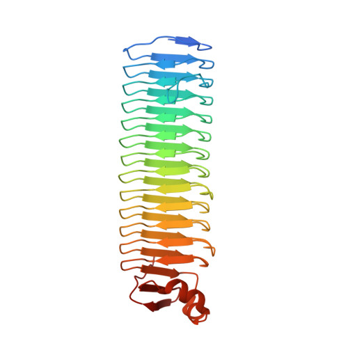
    The mechanism by which antifreeze proteins (AFPs) irreversibly bind to ice has not yet been resolved. The ice-binding site of an AFP is relatively hydrophobic, but also contains many potential hydrogen bond donors/acceptors. The extent to which hydrogen bonding and the hydrophobic effect contribute to ice binding has been debated for over 30 years. Here we have elucidated the ice-binding mechanism through solving the first crystal structure of an Antarctic bacterial AFP. This 34-kDa domain, the largest AFP structure determined to date, folds as a Ca(2+)-bound parallel beta-helix with an extensive array of ice-like surface waters that are anchored via hydrogen bonds directly to the polypeptide backbone and adjacent side chains. These bound waters make an excellent three-dimensional match to both the primary prism and basal planes of ice and in effect provide an extensive X-ray crystallographic picture of the AFPice interaction. This unobstructed view, free from crystal-packing artefacts, shows the contributions of both the hydrophobic effect and hydrogen bonding during AFP adsorption to ice. We term this mode of binding the "anchored clathrate" mechanism of AFP action.


  • Organizational Affiliation
    • Department of Biochemistry, Queen's University, Kingston, ON, Canada K7L 3N6.

Macromolecule Content 

  • Total Structure Weight: 135.73 kDa 
  • Atom Count: 10,219 
  • Modeled Residue Count: 1,227 
  • Deposited Residue Count: 1,292 
  • Unique protein chains: 1

Macromolecules

Find similar proteins by:|  3D Structure
Entity ID: 1
MoleculeChains  Sequence LengthOrganismDetailsImage
Antifreeze protein
A, B, C, D
323Marinomonas primoryensisMutation(s): 0 
Gene Names: MpAFP
UniProt
Find proteins for A1YIY3 (Marinomonas primoryensis)
Explore A1YIY3 
Go to UniProtKB:  A1YIY3
Entity Groups
Sequence Clusters30% Identity50% Identity70% Identity90% Identity95% Identity100% Identity
UniProt GroupA1YIY3
Sequence Annotations
Expand
Reference Sequence

Small Molecules

Ligands 3 Unique
IDChains Name / Formula / InChI Key2D Diagram3D Interactions
ACT

Query on ACT


Download:Ideal Coordinates CCD File
IA [auth B],
WA [auth C]
ACETATE ION
C2 H3 O2
QTBSBXVTEAMEQO-UHFFFAOYSA-M
CA

Query on CA


Download:Ideal Coordinates CCD File
AA [auth B]
AB [auth D]
BA [auth B]
BB [auth D]
CA [auth B]
AA [auth B],
AB [auth D],
BA [auth B],
BB [auth D],
CA [auth B],
CB [auth D],
DA [auth B],
DB [auth D],
E [auth A],
EA [auth B],
EB [auth D],
F [auth A],
FA [auth B],
FB [auth D],
G [auth A],
GA [auth B],
GB [auth D],
H [auth A],
HB [auth D],
I [auth A],
IB [auth D],
J [auth A],
JA [auth C],
JB [auth D],
K [auth A],
KA [auth C],
L [auth A],
LA [auth C],
M [auth A],
MA [auth C],
N [auth A],
NA [auth C],
O [auth A],
OA [auth C],
P [auth A],
PA [auth C],
Q [auth A],
QA [auth C],
RA [auth C],
SA [auth C],
TA [auth C],
U [auth B],
UA [auth C],
V [auth B],
VA [auth C],
W [auth B],
X [auth B],
XA [auth D],
Y [auth B],
YA [auth D],
Z [auth B],
ZA [auth D]
CALCIUM ION
Ca
BHPQYMZQTOCNFJ-UHFFFAOYSA-N
MG

Query on MG


Download:Ideal Coordinates CCD File
HA [auth B],
R [auth A],
S [auth A],
T [auth A]
MAGNESIUM ION
Mg
JLVVSXFLKOJNIY-UHFFFAOYSA-N

Experimental Data & Validation

Experimental Data

  • Method: X-RAY DIFFRACTION
  • Resolution: 1.70 Å
  • R-Value Free:  0.200 (Depositor), 0.214 (DCC) 
  • R-Value Work:  0.164 (Depositor) 
  • R-Value Observed: 0.165 (Depositor) 
Space Group: P 1
Unit Cell:
Length ( Å )Angle ( ˚ )
a = 57.5α = 99.06
b = 70.78β = 92.3
c = 84.25γ = 97.47
Software Package:
Software NamePurpose
HKL-2000data collection
SHELXSphasing
REFMACrefinement
HKL-2000data reduction
SCALAdata scaling

Structure Validation

View Full Validation Report


Entry History 

Deposition Data

Revision History  (Full details and data files)

  • Version 1.0: 2011-04-27
    Type: Initial release
  • Version 1.1: 2011-07-13
    Changes: Version format compliance
  • Version 1.2: 2024-02-21
    Changes: Data collection, Database references, Derived calculations