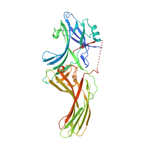
3P2D

Crystal structure of arrestin-3 reveals the basis of the difference in receptor binding between two non-visual subtypes


Experimental Data Snapshot

  • Method: X-RAY DIFFRACTION
  • Resolution: 3.00 Å
  • R-Value Free: 0.286 
  • R-Value Work: 0.219 
  • R-Value Observed: 0.223 

Starting Model: experimental
View more details

wwPDB Validation   3D Report Full Report


This is version 1.2 of the entry. See complete history


Literature

Crystal Structure of Arrestin-3 Reveals the Basis of the Difference in Receptor Binding Between Two Non-visual Subtypes.

Zhan, X.Gimenez, L.E.Gurevich, V.V.Spiller, B.W.

(2011) J Mol Biol 406: 467-478

  • DOI: https://doi.org/10.1016/j.jmb.2010.12.034
  • Primary Citation of Related Structures:  
    3P2D

  • PubMed Abstract: 

    Arrestins are multi-functional proteins that regulate signaling and trafficking of the majority of G protein-coupled receptors (GPCRs), as well as sub-cellular localization and activity of many other signaling proteins. We report the first crystal structure of arrestin-3, solved at 3.0 Å resolution. Arrestin-3 is an elongated two-domain molecule with overall fold and key inter-domain interactions that hold the free protein in the basal conformation similar to the other subtypes. Arrestin-3 is the least selective member of the family, binding a wide variety of GPCRs with high affinity and demonstrating lower preference for active phosphorylated forms of the receptors. In contrast to the other three arrestins, part of the receptor-binding surface in the arrestin-3 C-domain does not form a contiguous β-sheet, which is consistent with increased flexibility. By swapping the corresponding elements between arrestin-2 and arrestin-3 we show that the presence of this loose structure is correlated with reduced arrestin selectivity for activated receptors, consistent with a conformational change in this β-sheet upon receptor binding.


  • Organizational Affiliation

    Department of Pharmacology, Vanderbilt University, Nashville, TN 37232, USA.


Macromolecules
Find similar proteins by:  (by identity cutoff)  |  3D Structure
Entity ID: 1
MoleculeChains Sequence LengthOrganismDetailsImage
Beta-arrestin-2
A, B
393Bos taurusMutation(s): 0 
Gene Names: ARRB2
UniProt
Find proteins for P32120 (Bos taurus)
Explore P32120 
Go to UniProtKB:  P32120
Entity Groups  
Sequence Clusters30% Identity50% Identity70% Identity90% Identity95% Identity100% Identity
UniProt GroupP32120
Sequence Annotations
Expand
  • Reference Sequence
Experimental Data & Validation

Experimental Data

  • Method: X-RAY DIFFRACTION
  • Resolution: 3.00 Å
  • R-Value Free: 0.286 
  • R-Value Work: 0.219 
  • R-Value Observed: 0.223 
  • Space Group: P 21 21 21
Unit Cell:
Length ( Å )Angle ( ˚ )
a = 73.177α = 90
b = 73.323β = 90
c = 201.973γ = 90
Software Package:
Software NamePurpose
ADSCdata collection
PHASERphasing
PHENIXrefinement
HKL-2000data reduction
HKL-2000data scaling

Structure Validation

View Full Validation Report


Entry History 

Deposition Data

Revision History  (Full details and data files)

  • Version 1.0: 2011-01-26
    Type: Initial release
  • Version 1.1: 2011-07-13
    Changes: Version format compliance
  • Version 1.2: 2023-09-06
    Changes: Data collection, Database references, Refinement description