3OXX | pdb_00003oxx

Crystal Structure of HIV-1 I50V, A71V Protease in Complex with the Protease Inhibitor Atazanavir


Experimental Data Snapshot

  • Method: X-RAY DIFFRACTION
  • Resolution: 1.65 Å
  • R-Value Free: 
    0.215 (Depositor), 0.228 (DCC) 
  • R-Value Work: 
    0.173 (Depositor), 0.187 (DCC) 
  • R-Value Observed: 
    0.175 (Depositor) 

Starting Model: experimental
View more details

wwPDB Validation 3D Report Full Report

Validation slider image for 3OXX

Ligand Structure Quality Assessment 


This is version 1.3 of the entry. See complete history

Literature

Structural and thermodynamic basis of amprenavir/darunavir and atazanavir resistance in HIV-1 protease with mutations at residue 50.

Mittal, S.Bandaranayake, R.M.King, N.M.Prabu-Jeyabalan, M.Nalam, M.N.Nalivaika, E.A.Yilmaz, N.K.Schiffer, C.A.

(2013) J Virol 87: 4176-4184

  • DOI: https://doi.org/10.1128/JVI.03486-12
  • Primary Citation Related Structures: 
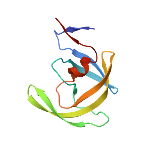
    3OXV, 3OXW, 3OXX

  • PubMed Abstract: 

    Drug resistance occurs through a series of subtle changes that maintain substrate recognition but no longer permit inhibitor binding. In HIV-1 protease, mutations at I50 are associated with such subtle changes that confer differential resistance to specific inhibitors. Residue I50 is located at the protease flap tips, closing the active site upon ligand binding. Under selective drug pressure, I50V/L substitutions emerge in patients, compromising drug susceptibility and leading to treatment failure. The I50V substitution is often associated with amprenavir (APV) and darunavir (DRV) resistance, while the I50L substitution is observed in patients failing atazanavir (ATV) therapy. To explain how APV, DRV, and ATV susceptibility are influenced by mutations at residue 50 in HIV-1 protease, structural and binding thermodynamics studies were carried out on I50V/L-substituted protease variants in the compensatory mutation A71V background. Reduced affinity to both I50V/A71V and I50L/A71V double mutants is largely due to decreased binding entropy, which is compensated for by enhanced enthalpy for ATV binding to I50V variants and APV binding to I50L variants, leading to hypersusceptibility in these two cases. Analysis of the crystal structures showed that the substitutions at residue 50 affect how APV, DRV, and ATV bind the protease with altered van der Waals interactions and that the selection of I50V versus I50L is greatly influenced by the chemical moieties at the P1 position for APV/DRV and the P2 position for ATV. Thus, the varied inhibitor susceptibilities of I50V/L protease variants are largely a direct consequence of the interdependent changes in protease inhibitor interactions.


  • Organizational Affiliation
    • Department of Biochemistry and Molecular Pharmacology, University of Massachusetts Medical School, Worcester, Massachusetts, USA.

Macromolecule Content 

  • Total Structure Weight: 45.86 kDa 
  • Atom Count: 3,285 
  • Modeled Residue Count: 396 
  • Deposited Residue Count: 396 
  • Unique protein chains: 1

Macromolecules

Find similar proteins by:|  3D Structure
Entity ID: 1
MoleculeChains  Sequence LengthOrganismDetailsImage
HIV-1 PROTEASEA [auth B],
B [auth A],
C,
D
99HIV-1 M:B_ARV2/SF2Mutation(s): 3 
Gene Names: gag-pol
EC: 3.4.23.16
UniProt
Find proteins for P03369 (Human immunodeficiency virus type 1 group M subtype B (isolate ARV2/SF2))
Explore P03369 
Go to UniProtKB:  P03369
Entity Groups
Sequence Clusters30% Identity50% Identity70% Identity90% Identity95% Identity100% Identity
UniProt GroupP03369
Sequence Annotations
Expand
Reference Sequence

Small Molecules

Ligands 5 Unique
IDChains Name / Formula / InChI Key2D Diagram3D Interactions
DR7

Query on DR7


Download:Ideal Coordinates CCD File
K [auth A],
N [auth C]
(3S,8S,9S,12S)-3,12-BIS(1,1-DIMETHYLETHYL)-8-HYDROXY-4,11-DIOXO-9-(PHENYLMETHYL)-6-[[4-(2-PYRIDINYL)PHENYL]METHYL]-2,5, 6,10,13-PENTAAZATETRADECANEDIOIC ACID DIMETHYL ESTER
C38 H52 N6 O7
AXRYRYVKAWYZBR-GASGPIRDSA-N
PO4

Query on PO4


Download:Ideal Coordinates CCD File
T [auth D]PHOSPHATE ION
O4 P
NBIIXXVUZAFLBC-UHFFFAOYSA-K
GOL

Query on GOL


Download:Ideal Coordinates CCD File
E [auth B]
F [auth B]
L [auth A]
O [auth C]
P [auth C]
E [auth B],
F [auth B],
L [auth A],
O [auth C],
P [auth C],
S [auth D]
GLYCEROL
C3 H8 O3
PEDCQBHIVMGVHV-UHFFFAOYSA-N
EDO

Query on EDO


Download:Ideal Coordinates CCD File
G [auth B],
M [auth A],
V [auth D]
1,2-ETHANEDIOL
C2 H6 O2
LYCAIKOWRPUZTN-UHFFFAOYSA-N
ACT

Query on ACT


Download:Ideal Coordinates CCD File
H [auth B]
I [auth B]
J [auth B]
Q [auth C]
R [auth C]
H [auth B],
I [auth B],
J [auth B],
Q [auth C],
R [auth C],
U [auth D]
ACETATE ION
C2 H3 O2
QTBSBXVTEAMEQO-UHFFFAOYSA-M

Experimental Data & Validation

Experimental Data

  • Method: X-RAY DIFFRACTION
  • Resolution: 1.65 Å
  • R-Value Free:  0.215 (Depositor), 0.228 (DCC) 
  • R-Value Work:  0.173 (Depositor), 0.187 (DCC) 
  • R-Value Observed: 0.175 (Depositor) 
Space Group: P 1 21 1
Unit Cell:
Length ( Å )Angle ( ˚ )
a = 51.238α = 90
b = 58.489β = 95.88
c = 62.118γ = 90
Software Package:
Software NamePurpose
DENZOdata reduction
SCALEPACKdata scaling
PHASERphasing
REFMACrefinement
PDB_EXTRACTdata extraction
HKL-2000data reduction
HKL-2000data scaling

Structure Validation

View Full Validation Report


Ligand Structure Quality Assessment 


Entry History 

Deposition Data

Revision History  (Full details and data files)

  • Version 1.0: 2011-09-28
    Type: Initial release
  • Version 1.1: 2015-04-01
    Changes: Database references
  • Version 1.2: 2017-11-08
    Changes: Refinement description
  • Version 1.3: 2023-09-06
    Changes: Data collection, Database references, Derived calculations, Refinement description, Structure summary