3OUF | pdb_00003ouf

Structure of a K+ selective NaK mutant


Experimental Data Snapshot

  • Method: X-RAY DIFFRACTION
  • Resolution: 1.55 Å
  • R-Value Free: 
    0.215 (Depositor), 0.214 (DCC) 
  • R-Value Work: 
    0.200 (Depositor), 0.215 (DCC) 

wwPDB Validation 3D Report Full Report

Validation slider image for 3OUF

This is version 1.3 of the entry. See complete history

Literature

Tuning the ion selectivity of tetrameric cation channels by changing the number of ion binding sites.

Derebe, M.G.Sauer, D.B.Zeng, W.Alam, A.Shi, N.Jiang, Y.

(2011) Proc Natl Acad Sci U S A 108: 598-602

  • DOI: https://doi.org/10.1073/pnas.1013636108
  • Primary Citation Related Structures: 
    3K03, 3OUF, 3OUS

  • PubMed Abstract: 

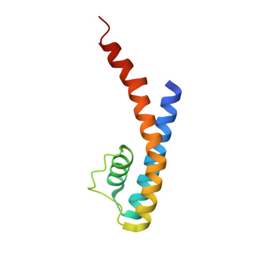
    Selective ion conduction across ion channel pores is central to cellular physiology. To understand the underlying principles of ion selectivity in tetrameric cation channels, we engineered a set of cation channel pores based on the nonselective NaK channel and determined their structures to high resolution. These structures showcase an ensemble of selectivity filters with a various number of contiguous ion binding sites ranging from 2 to 4, with each individual site maintaining a geometry and ligand environment virtually identical to that of equivalent sites in K(+) channel selectivity filters. Combined with single channel electrophysiology, we show that only the channel with four ion binding sites is K(+) selective, whereas those with two or three are nonselective and permeate Na(+) and K(+) equally well. These observations strongly suggest that the number of contiguous ion binding sites in a single file is the key determinant of the channel's selectivity properties and the presence of four sites in K(+) channels is essential for highly selective and efficient permeation of K(+) ions.


  • Organizational Affiliation
    • Department of Physiology, University of Texas Southwestern Medical Center, Dallas, TX 75390-9040, USA.

Macromolecule Content 

  • Total Structure Weight: 22.49 kDa 
  • Atom Count: 1,607 
  • Modeled Residue Count: 189 
  • Deposited Residue Count: 194 
  • Unique protein chains: 1

Macromolecules

Find similar proteins by:|  3D Structure
Entity ID: 1
MoleculeChains  Sequence LengthOrganismDetailsImage
Potassium channel protein
A, B
97Bacillus cereus m1550Mutation(s): 1 
Gene Names: bcere0011_5790

Small Molecules

Ligands 2 Unique
IDChains Name / Formula / InChI Key2D Diagram3D Interactions
MPD

Query on MPD


Download:Ideal Coordinates CCD File
C [auth A],
D [auth A],
J [auth B],
K [auth B],
L [auth B]
(4S)-2-METHYL-2,4-PENTANEDIOL
C6 H14 O2
SVTBMSDMJJWYQN-YFKPBYRVSA-N
K

Query on K


Download:Ideal Coordinates CCD File
E [auth A]
F [auth A]
G [auth A]
H [auth A]
I [auth A]
E [auth A],
F [auth A],
G [auth A],
H [auth A],
I [auth A],
M [auth B],
N [auth B],
O [auth B],
P [auth B]
POTASSIUM ION
K
NPYPAHLBTDXSSS-UHFFFAOYSA-N

Experimental Data & Validation

Experimental Data

  • Method: X-RAY DIFFRACTION
  • Resolution: 1.55 Å
  • R-Value Free:  0.215 (Depositor), 0.214 (DCC) 
  • R-Value Work:  0.200 (Depositor), 0.215 (DCC) 
Space Group: I 4
Unit Cell:
Length ( Å )Angle ( ˚ )
a = 67.939α = 90
b = 67.939β = 90
c = 89.638γ = 90
Software Package:
Software NamePurpose
DENZOdata reduction
SCALEPACKdata scaling
CNSrefinement
PDB_EXTRACTdata extraction
HKL-2000data collection
CNSphasing

Structure Validation

View Full Validation Report


Entry History 

Deposition Data

Revision History  (Full details and data files)

  • Version 1.0: 2011-01-12
    Type: Initial release
  • Version 1.1: 2011-07-13
    Changes: Version format compliance
  • Version 1.2: 2017-11-08
    Changes: Refinement description
  • Version 1.3: 2024-02-21
    Changes: Data collection, Database references, Derived calculations