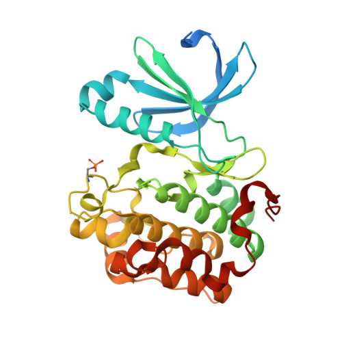
3ORX

PDK1 mutant bound to allosteric disulfide fragment inhibitor 1F8


Experimental Data Snapshot

  • Method: X-RAY DIFFRACTION
  • Resolution: 2.20 Å
  • R-Value Free: 0.243 
  • R-Value Work: 0.208 
  • R-Value Observed: 0.209 

Starting Model: experimental
View more details

wwPDB Validation   3D Report Full Report


Ligand Structure Quality Assessment 


This is version 1.3 of the entry. See complete history


Literature

Turning a protein kinase on or off from a single allosteric site via disulfide trapping.

Sadowsky, J.D.Burlingame, M.A.Wolan, D.W.McClendon, C.L.Jacobson, M.P.Wells, J.A.

(2011) Proc Natl Acad Sci U S A 108: 6056-6061

  • DOI: https://doi.org/10.1073/pnas.1102376108
  • Primary Citation of Related Structures:  
    3ORX, 3ORZ, 3OTU

  • PubMed Abstract: 

    There is significant interest in identifying and characterizing allosteric sites in enzymes such as protein kinases both for understanding allosteric mechanisms as well as for drug discovery. Here, we apply a site-directed technology, disulfide trapping, to interrogate structurally and functionally how an allosteric site on the Ser/Thr kinase, 3-phosphoinositide-dependent kinase 1 (PDK1)--the PDK1-interacting-fragment (PIF) pocket--is engaged by an activating peptide motif on downstream substrate kinases (PIFtides) and by small molecule fragments. By monitoring pairwise disulfide conjugation between PIFtide and PDK1 cysteine mutants, we defined the PIFtide binding orientation in the PIF pocket of PDK1 and assessed subtle relationships between PIFtide positioning and kinase activation. We also discovered a variety of small molecule fragment disulfides (< 300 Da) that could either activate or inhibit PDK1 by conjugation to the PIF pocket, thus displaying greater functional diversity than is displayed by PIFtides conjugated to the same sites. Biochemical data and three crystal structures provided insight into the mechanism of action of the best fragment activators and inhibitors. These studies show that disulfide trapping is useful for characterizing allosteric sites on kinases and that a single allosteric site on a protein kinase can be exploited for both activation and inhibition by small molecules.


  • Organizational Affiliation

    Departments of Pharmaceutical Chemistry and Cellular and Molecular Pharmacology, University of California, 1700 Fourth Street, San Francisco, CA 94158, USA.


Macromolecules
Find similar proteins by:  (by identity cutoff)  |  3D Structure
Entity ID: 1
MoleculeChains Sequence LengthOrganismDetailsImage
3-phosphoinositide-dependent protein kinase 1
A, B, C, D, E
A, B, C, D, E, F, G, H
316Homo sapiensMutation(s): 1 
Gene Names: PDK1PDPK1
EC: 2.7.11.1
UniProt & NIH Common Fund Data Resources
Find proteins for O15530 (Homo sapiens)
Explore O15530 
Go to UniProtKB:  O15530
PHAROS:  O15530
GTEx:  ENSG00000140992 
Entity Groups  
Sequence Clusters30% Identity50% Identity70% Identity90% Identity95% Identity100% Identity
UniProt GroupO15530
Sequence Annotations
Expand
  • Reference Sequence
Small Molecules
Ligands 2 Unique
IDChains Name / Formula / InChI Key2D Diagram3D Interactions
1F8
Query on 1F8

Download Ideal Coordinates CCD File 
I [auth A]
K [auth B]
M [auth C]
O [auth D]
Q [auth E]
I [auth A],
K [auth B],
M [auth C],
O [auth D],
Q [auth E],
R [auth F],
S [auth G],
T [auth H]
2-methyl-N-(2-sulfanylethyl)-1-benzofuran-3-carboxamide
C12 H13 N O2 S
MMIVKJYKVVAJCP-UHFFFAOYSA-N
CL
Query on CL

Download Ideal Coordinates CCD File 
J [auth A],
L [auth B],
N [auth C],
P [auth D]
CHLORIDE ION
Cl
VEXZGXHMUGYJMC-UHFFFAOYSA-M
Modified Residues  1 Unique
IDChains TypeFormula2D DiagramParent
SEP
Query on SEP
A, B, C, D, E
A, B, C, D, E, F, G, H
L-PEPTIDE LINKINGC3 H8 N O6 PSER
Experimental Data & Validation

Experimental Data

  • Method: X-RAY DIFFRACTION
  • Resolution: 2.20 Å
  • R-Value Free: 0.243 
  • R-Value Work: 0.208 
  • R-Value Observed: 0.209 
  • Space Group: P 1
Unit Cell:
Length ( Å )Angle ( ˚ )
a = 40.289α = 91.76
b = 115.87β = 89.99
c = 145.718γ = 95.41
Software Package:
Software NamePurpose
CCP4model building
PHENIXrefinement
Blu-Icedata collection
HKL-2000data reduction
HKL-2000data scaling
CCP4phasing

Structure Validation

View Full Validation Report


Ligand Structure Quality Assessment 


Entry History 

Deposition Data

Revision History  (Full details and data files)

  • Version 1.0: 2011-03-23
    Type: Initial release
  • Version 1.1: 2011-07-13
    Changes: Version format compliance
  • Version 1.2: 2023-09-06
    Changes: Data collection, Database references, Derived calculations, Refinement description
  • Version 1.3: 2024-11-20
    Changes: Structure summary