3OM5 | pdb_00003om5

Crystal structure of B. megaterium levansucrase mutant N252A


Experimental Data Snapshot

  • Method: X-RAY DIFFRACTION
  • Resolution: 1.95 Å
  • R-Value Free: 
    0.208 (Depositor), 0.235 (DCC) 
  • R-Value Work: 
    0.181 (Depositor), 0.205 (DCC) 
  • R-Value Observed: 
    0.182 (Depositor) 

wwPDB Validation 3D Report Full Report

Validation slider image for 3OM5

This is version 1.2 of the entry. See complete history

Literature

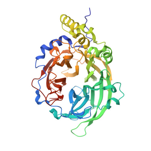
Polysaccharide Synthesis of the Levansucrase SacB from Bacillus megaterium Is Controlled by Distinct Surface Motifs.

Strube, C.P.Homann, A.Gamer, M.Jahn, D.Seibel, J.Heinz, D.W.

(2011) J Biological Chem 286: 17593-17600

  • DOI: https://doi.org/10.1074/jbc.M110.203166
  • Primary Citation Related Structures: 
    3OM2, 3OM5, 3OM6, 3OM7

  • PubMed Abstract: 

    Despite the widespread biological function of carbohydrates, the polysaccharide synthesis mechanisms of glycosyltransferases remain largely unexplored. Bacterial levansucrases (glycoside hydrolase family 68) synthesize high molecular weight, β-(2,6)-linked levan from sucrose by transfer of fructosyl units. The kinetic and biochemical characterization of Bacillus megaterium levansucrase SacB variants Y247A, Y247W, N252A, D257A, and K373A reveal novel surface motifs remote from the sucrose binding site with distinct influence on the polysaccharide product spectrum. The wild type activity (k(cat)) and substrate affinity (K(m)) are maintained. The structures of the SacB variants reveal clearly distinguishable subsites for polysaccharide synthesis as well as an intact active site architecture. These results lead to a new understanding of polysaccharide synthesis mechanisms. The identified surface motifs are discussed in the context of related glycosyltransferases.


  • Organizational Affiliation
    • Department of Molecular Structural Biology, Helmholtz-Centre for Infection Research, Inhoffenstrasse 7B, 38124 Braunschweig, Germany.

Macromolecule Content 

  • Total Structure Weight: 205.84 kDa 
  • Atom Count: 15,439 
  • Modeled Residue Count: 1,792 
  • Deposited Residue Count: 1,824 
  • Unique protein chains: 1

Macromolecules

Find similar proteins by:|  3D Structure
Entity ID: 1
MoleculeChains  Sequence LengthOrganismDetailsImage
Levansucrase
A, B, C, D
456Priestia megateriumMutation(s): 1 
Gene Names: sacB
EC: 2.4.1.10
UniProt
Find proteins for D5DC07 (Priestia megaterium (strain DSM 319 / IMG 1521))
Explore D5DC07 
Go to UniProtKB:  D5DC07
Entity Groups
Sequence Clusters30% Identity50% Identity70% Identity90% Identity95% Identity100% Identity
UniProt GroupD5DC07
Sequence Annotations
Expand
Reference Sequence

Small Molecules

Ligands 5 Unique
IDChains Name / Formula / InChI Key2D Diagram3D Interactions
P6G

Query on P6G


Download:Ideal Coordinates CCD File
F [auth A],
H [auth B]
HEXAETHYLENE GLYCOL
C12 H26 O7
IIRDTKBZINWQAW-UHFFFAOYSA-N
PGE

Query on PGE


Download:Ideal Coordinates CCD File
J [auth C],
K [auth C]
TRIETHYLENE GLYCOL
C6 H14 O4
ZIBGPFATKBEMQZ-UHFFFAOYSA-N
PEG

Query on PEG


Download:Ideal Coordinates CCD File
L [auth C],
P [auth D]
DI(HYDROXYETHYL)ETHER
C4 H10 O3
MTHSVFCYNBDYFN-UHFFFAOYSA-N
SO4

Query on SO4


Download:Ideal Coordinates CCD File
M [auth C],
N [auth C],
Q [auth D],
R [auth D],
S [auth D]
SULFATE ION
O4 S
QAOWNCQODCNURD-UHFFFAOYSA-L
CA

Query on CA


Download:Ideal Coordinates CCD File
E [auth A],
G [auth B],
I [auth C],
O [auth D]
CALCIUM ION
Ca
BHPQYMZQTOCNFJ-UHFFFAOYSA-N

Experimental Data & Validation

Experimental Data

  • Method: X-RAY DIFFRACTION
  • Resolution: 1.95 Å
  • R-Value Free:  0.208 (Depositor), 0.235 (DCC) 
  • R-Value Work:  0.181 (Depositor), 0.205 (DCC) 
  • R-Value Observed: 0.182 (Depositor) 
Space Group: P 1 21 1
Unit Cell:
Length ( Å )Angle ( ˚ )
a = 93.368α = 90
b = 100.186β = 90.68
c = 95.195γ = 90
Software Package:
Software NamePurpose
REFMACrefinement
PDB_EXTRACTdata extraction
DNAdata collection
XDSdata reduction
SCALAdata scaling
MOLREPphasing

Structure Validation

View Full Validation Report


Entry History 

Deposition Data

Revision History  (Full details and data files)

  • Version 1.0: 2011-03-23
    Type: Initial release
  • Version 1.1: 2011-07-13
    Changes: Version format compliance
  • Version 1.2: 2024-02-21
    Changes: Data collection, Database references, Derived calculations