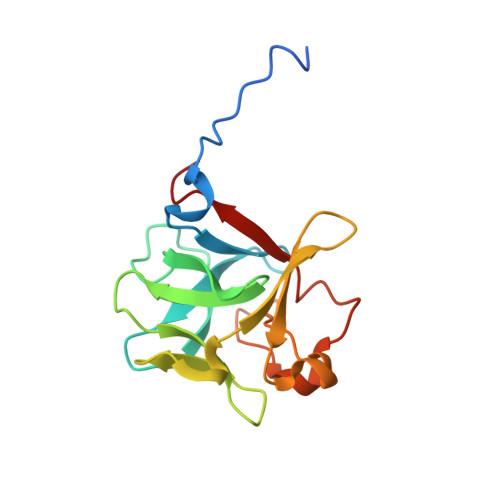
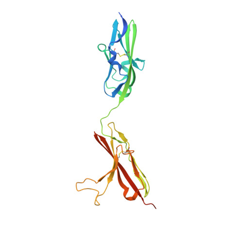
3OJM

Crystal Structure of FGF1 complexed with the ectodomain of FGFR2b harboring P253R Apert mutation


Experimental Data Snapshot

  • Method: X-RAY DIFFRACTION
  • Resolution: 2.10 Å
  • R-Value Free: 0.263 
  • R-Value Work: 0.229 

Starting Model: experimental
View more details

wwPDB Validation   3D Report Full Report


This is version 1.3 of the entry. See complete history


Literature

Plasticity in Interactions of Fibroblast Growth Factor 1 (FGF1) N Terminus with FGF Receptors Underlies Promiscuity of FGF1.

Beenken, A.Eliseenkova, A.V.Ibrahimi, O.A.Olsen, S.K.Mohammadi, M.

(2012) J Biol Chem 287: 3067-3078

  • DOI: https://doi.org/10.1074/jbc.M111.275891
  • Primary Citation of Related Structures:  
    3OJ2, 3OJM, 3OJV

  • PubMed Abstract: 

    Tissue-specific alternative splicing in the second half of Ig-like domain 3 (D3) of fibroblast growth factor receptors 1-3 (FGFR1 to -3) generates epithelial FGFR1b-FGFR3b and mesenchymal FGFR1c-FGFR3c splice isoforms. This splicing event establishes a selectivity filter to restrict the ligand binding specificity of FGFRb and FGFRc isoforms to mesenchymally and epithelially derived fibroblast growth factors (FGFs), respectively. FGF1 is termed the "universal FGFR ligand" because it overrides this specificity barrier. To elucidate the molecular basis for FGF1 cross-reactivity with the "b" and "c" splice isoforms of FGFRs, we determined the first crystal structure of FGF1 in complex with an FGFRb isoform, FGFR2b, at 2.1 Å resolution. Comparison of the FGF1-FGFR2b structure with the three previously published FGF1-FGFRc structures reveals that plasticity in the interactions of the N-terminal region of FGF1 with FGFR D3 is the main determinant of FGF1 cross-reactivity with both isoforms of FGFRs. In support of our structural data, we demonstrate that substitution of three N-terminal residues (Gly-19, His-25, and Phe-26) of FGF2 (a ligand that does not bind FGFR2b) for the corresponding residues of FGF1 (Phe-16, Asn-22, and Tyr-23) enables the FGF2 triple mutant to bind and activate FGFR2b. These findings taken together with our previous structural data on receptor binding specificity of FGF2, FGF8, and FGF10 conclusively show that sequence divergence at the N termini of FGFs is the primary regulator of the receptor binding specificity and promiscuity of FGFs.


  • Organizational Affiliation

    Department of Pharmacology, New York University School of Medicine, New York, New York 10016, USA.


Macromolecules
Find similar proteins by:  (by identity cutoff)  |  3D Structure
Entity ID: 1
MoleculeChains Sequence LengthOrganismDetailsImage
Heparin-binding growth factor 1155Homo sapiensMutation(s): 0 
Gene Names: FGF1FGFA
UniProt & NIH Common Fund Data Resources
Find proteins for P05230 (Homo sapiens)
Explore P05230 
Go to UniProtKB:  P05230
PHAROS:  P05230
GTEx:  ENSG00000113578 
Entity Groups  
Sequence Clusters30% Identity50% Identity70% Identity90% Identity95% Identity100% Identity
UniProt GroupP05230
Sequence Annotations
Expand
  • Reference Sequence
Find similar proteins by:  (by identity cutoff)  |  3D Structure
Entity ID: 2
MoleculeChains Sequence LengthOrganismDetailsImage
Fibroblast growth factor receptor 2231Homo sapiensMutation(s): 1 
Gene Names: BEKFGFR2KGFRKSAM
EC: 2.7.10.1
UniProt & NIH Common Fund Data Resources
Find proteins for P21802 (Homo sapiens)
Explore P21802 
Go to UniProtKB:  P21802
PHAROS:  P21802
GTEx:  ENSG00000066468 
Entity Groups  
Sequence Clusters30% Identity50% Identity70% Identity90% Identity95% Identity100% Identity
UniProt GroupP21802
Sequence Annotations
Expand
  • Reference Sequence
Experimental Data & Validation

Experimental Data

  • Method: X-RAY DIFFRACTION
  • Resolution: 2.10 Å
  • R-Value Free: 0.263 
  • R-Value Work: 0.229 
  • Space Group: P 21 21 21
Unit Cell:
Length ( Å )Angle ( ˚ )
a = 66.74α = 90
b = 72.349β = 90
c = 91.912γ = 90
Software Package:
Software NamePurpose
HKL-2000data collection
AMoREphasing
CNSrefinement
HKL-2000data reduction
HKL-2000data scaling

Structure Validation

View Full Validation Report


Entry History 

Deposition Data

Revision History  (Full details and data files)

  • Version 1.0: 2011-08-31
    Type: Initial release
  • Version 1.1: 2012-03-07
    Changes: Database references
  • Version 1.2: 2023-09-06
    Changes: Data collection, Database references, Derived calculations, Refinement description
  • Version 1.3: 2024-11-06
    Changes: Structure summary