3OG6

The crystal structure of human interferon lambda 1 complexed with its high affinity receptor in space group P212121


Experimental Data Snapshot

  • Method: X-RAY DIFFRACTION
  • Resolution: 2.10 Å
  • R-Value Free: 0.228 
  • R-Value Work: 0.184 
  • R-Value Observed: 0.185 

Starting Models: experimental
View more details

wwPDB Validation   3D Report Full Report


Ligand Structure Quality Assessment 


This is version 2.1 of the entry. See complete history


Literature

Crystal structure of the complex of human interferon-lambda1 with its high affinity receptor interferon-lambdaR1.

Miknis, Z.J.Magracheva, E.Li, W.Zdanov, A.Kotenko, S.V.Wlodawer, A.

(2010) J Mol Biol 404: 650-664

  • DOI: https://doi.org/10.1016/j.jmb.2010.09.068
  • Primary Citation of Related Structures:  
    3OG4, 3OG6

  • PubMed Abstract: 

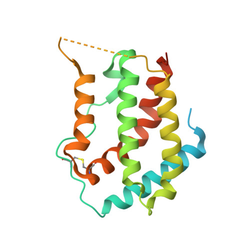
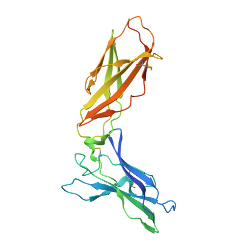
    Interferon (IFN)-λ1 [also known as interleukin (IL)-29] belongs to the recently discovered group of type III IFNs. All type III IFNs initiate signaling processes through formation of specific heterodimeric receptor complexes consisting of IFN-λR1 and IL-10R2. We have determined the structure of human IFN-λ1 complexed with human IFN-λR1, a receptor unique to type III IFNs. The overall structure of IFN-λ1 is topologically similar to the structure of IL-10 and other members of the IL-10 family of cytokines. IFN-λR1 consists of two distinct domains having fibronectin type III topology. The ligand-receptor interface includes helix A, loop AB, and helix F on the IFN site, as well as loops primarily from the N-terminal domain and inter-domain hinge region of IFN-λR1. Composition and architecture of the interface that includes only a few direct hydrogen bonds support an idea that long-range ionic interactions between ligand and receptor govern the process of initial recognition of the molecules while hydrophobic interactions finalize it.


  • Organizational Affiliation

    Macromolecular Crystallography Laboratory, NCI-Frederick, Frederick, MD 21702, USA.


Macromolecules
Find similar proteins by:  (by identity cutoff)  |  3D Structure
Entity ID: 1
MoleculeChains Sequence LengthOrganismDetailsImage
Interleukin-29196Homo sapiensMutation(s): 2 
Gene Names: IFNL1IL29ZCYTO21
UniProt & NIH Common Fund Data Resources
Find proteins for Q8IU54 (Homo sapiens)
Explore Q8IU54 
Go to UniProtKB:  Q8IU54
GTEx:  ENSG00000182393 
Entity Groups  
Sequence Clusters30% Identity50% Identity70% Identity90% Identity95% Identity100% Identity
UniProt GroupQ8IU54
Sequence Annotations
Expand
  • Reference Sequence
Find similar proteins by:  (by identity cutoff)  |  3D Structure
Entity ID: 2
MoleculeChains Sequence LengthOrganismDetailsImage
Interleukin 28 receptor, alpha (Interferon, lambda receptor)226Homo sapiensMutation(s): 0 
Gene Names: hCG_1982865IL28RARP11-10N16.1-001
UniProt & NIH Common Fund Data Resources
Find proteins for Q8IU57 (Homo sapiens)
Explore Q8IU57 
Go to UniProtKB:  Q8IU57
PHAROS:  Q8IU57
GTEx:  ENSG00000185436 
Entity Groups  
Sequence Clusters30% Identity50% Identity70% Identity90% Identity95% Identity100% Identity
UniProt GroupQ8IU57
Glycosylation
Glycosylation Sites: 3Go to GlyGen: Q8IU57-1
Sequence Annotations
Expand
  • Reference Sequence
Oligosaccharides

Help

Entity ID: 3
MoleculeChains Length2D Diagram Glycosylation3D Interactions
beta-D-mannopyranose-(1-4)-2-acetamido-2-deoxy-beta-D-glucopyranose-(1-4)-2-acetamido-2-deoxy-beta-D-glucopyranose
C
3N-Glycosylation
Glycosylation Resources
GlyTouCan:  G15407YE
GlyCosmos:  G15407YE
GlyGen:  G15407YE
Experimental Data & Validation

Experimental Data

  • Method: X-RAY DIFFRACTION
  • Resolution: 2.10 Å
  • R-Value Free: 0.228 
  • R-Value Work: 0.184 
  • R-Value Observed: 0.185 
  • Space Group: P 21 21 21
Unit Cell:
Length ( Å )Angle ( ˚ )
a = 64.96α = 90
b = 85.79β = 90
c = 116.51γ = 90
Software Package:
Software NamePurpose
DENZOdata reduction
SCALEPACKdata scaling
PHASERphasing
PHENIXrefinement
PDB_EXTRACTdata extraction
HKL-2000data collection

Structure Validation

View Full Validation Report


Ligand Structure Quality Assessment 


Entry History 

Deposition Data

Revision History  (Full details and data files)

  • Version 1.0: 2010-10-20
    Type: Initial release
  • Version 1.1: 2011-07-13
    Changes: Version format compliance
  • Version 1.2: 2013-07-03
    Changes: Database references
  • Version 2.0: 2020-07-29
    Type: Remediation
    Reason: Carbohydrate remediation
    Changes: Atomic model, Data collection, Database references, Derived calculations, Structure summary
  • Version 2.1: 2023-09-06
    Changes: Data collection, Database references, Refinement description, Structure summary