


Crystal structure of the small GTPase RhebL1
Nedyalkova, L., Zhong, N., Tempel, W., Tong, Y., Shen, L., Loppnau, P., Arrowsmith, C.H., Edwards, A.M., Bountra, C., Weigelt, J., Bochkarev, A., Park, H.To be published.
Experimental Data Snapshot
Starting Model: experimental
View more details
Entity ID: 1 | |||||
|---|---|---|---|---|---|
| Molecule | Chains | Sequence Length | Organism | Details | Image |
| GTPase RhebL1 | 201 | Homo sapiens | Mutation(s): 0 Gene Names: RHEBL1 EC: 3.6.5 | 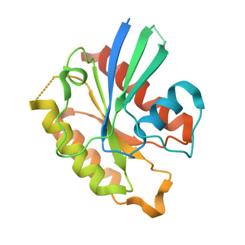 | |
UniProt & NIH Common Fund Data Resources | |||||
PHAROS: Q8TAI7 GTEx: ENSG00000167550 | |||||
Entity Groups | |||||
| Sequence Clusters | 30% Identity50% Identity70% Identity90% Identity95% Identity100% Identity | ||||
| UniProt Group | Q8TAI7 | ||||
Sequence AnnotationsExpand | |||||
Reference Sequence | |||||
| Ligands 2 Unique | |||||
|---|---|---|---|---|---|
| ID | Chains | Name / Formula / InChI Key | 2D Diagram | 3D Interactions | |
| GNP Download:Ideal Coordinates CCD File | C [auth A] | PHOSPHOAMINOPHOSPHONIC ACID-GUANYLATE ESTER C10 H17 N6 O13 P3 UQABYHGXWYXDTK-UUOKFMHZSA-N |  | ||
| MG Download:Ideal Coordinates CCD File | B [auth A] | MAGNESIUM ION Mg JLVVSXFLKOJNIY-UHFFFAOYSA-N |  | ||
| Length ( Å ) | Angle ( ˚ ) |
|---|---|
| a = 72.665 | α = 90 |
| b = 126.745 | β = 90 |
| c = 41.356 | γ = 90 |
| Software Name | Purpose |
|---|---|
| DENZO | data reduction |
| SCALEPACK | data scaling |
| PHASER | phasing |
| REFMAC | refinement |
| PDB_EXTRACT | data extraction |
| StructureStudio | data collection |
| HKL-2000 | data reduction |
| HKL-2000 | data scaling |


