3OEQ | pdb_00003oeq

Crystal structure of trimeric frataxin from the yeast Saccharomyces cerevisiae, with full length n-terminus


Experimental Data Snapshot

  • Method: X-RAY DIFFRACTION
  • Resolution: 2.96 Å
  • R-Value Free: 
    0.292 (Depositor), 0.291 (DCC) 
  • R-Value Work: 
    0.266 (Depositor), 0.261 (DCC) 
  • R-Value Observed: 
    0.268 (Depositor) 

Starting Model: experimental
View more details

wwPDB Validation 3D Report Full Report

Validation slider image for 3OEQ

This is version 1.3 of the entry. See complete history

Literature

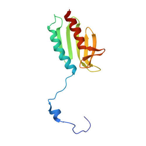
Oligomerization Propensity and Flexibility of Yeast Frataxin Studied by X-ray Crystallography and Small-Angle X-ray Scattering.

Soderberg, C.A.Shkumatov, A.V.Rajan, S.Gakh, O.Svergun, D.I.Isaya, G.Al-Karadaghi, S.

(2011) J Mol Biology 414: 783-797

  • DOI: https://doi.org/10.1016/j.jmb.2011.10.034
  • Primary Citation Related Structures: 
    3OEQ, 3OER

  • PubMed Abstract: 

    Frataxin is a mitochondrial protein with a central role in iron homeostasis. Defects in frataxin function lead to Friedreich's ataxia, a progressive neurodegenerative disease with childhood onset. The function of frataxin has been shown to be closely associated with its ability to form oligomeric species; however, the factors controlling oligomerization and the types of oligomers present in solution are a matter of debate. Using small-angle X-ray scattering, we found that Co(2+), glycerol, and a single amino acid substitution at the N-terminus, Y73A, facilitate oligomerization of yeast frataxin, resulting in a dynamic equilibrium between monomers, dimers, trimers, hexamers, and higher-order oligomers. Using X-ray crystallography, we found that Co(2+) binds inside the channel at the 3-fold axis of the trimer, which suggests that the metal has an oligomer-stabilizing role. The results reveal the types of oligomers present in solution and support our earlier suggestions that the trimer is the main building block of yeast frataxin oligomers. They also indicate that different mechanisms may control oligomer stability and oligomerization in vivo.


  • Organizational Affiliation
    • Center for Molecular Protein Science, Institute for Chemistry and Chemical Engineering, Lund University, SE-221 00 Lund, Sweden.

Macromolecule Content 

  • Total Structure Weight: 13.67 kDa 
  • Atom Count: 931 
  • Modeled Residue Count: 121 
  • Deposited Residue Count: 123 
  • Unique protein chains: 1

Macromolecules

Find similar proteins by:|  3D Structure
Entity ID: 1
MoleculeChains  Sequence LengthOrganismDetailsImage
Frataxin homolog, mitochondrial123Saccharomyces cerevisiaeMutation(s): 1 
Gene Names: YFH1YDL120W
EC: 1.16.3.1
UniProt
Find proteins for Q07540 (Saccharomyces cerevisiae (strain ATCC 204508 / S288c))
Explore Q07540 
Go to UniProtKB:  Q07540
Entity Groups
Sequence Clusters30% Identity50% Identity70% Identity90% Identity95% Identity100% Identity
UniProt GroupQ07540
Sequence Annotations
Expand
Reference Sequence

Experimental Data & Validation

Experimental Data

  • Method: X-RAY DIFFRACTION
  • Resolution: 2.96 Å
  • R-Value Free:  0.292 (Depositor), 0.291 (DCC) 
  • R-Value Work:  0.266 (Depositor), 0.261 (DCC) 
  • R-Value Observed: 0.268 (Depositor) 
Space Group: I 21 3
Unit Cell:
Length ( Å )Angle ( ˚ )
a = 121.36α = 90
b = 121.36β = 90
c = 121.36γ = 90
Software Package:
Software NamePurpose
MAR345dtbdata collection
AMoREphasing
REFMACrefinement
XDSdata reduction
XSCALEdata scaling

Structure Validation

View Full Validation Report


Entry History 

Deposition Data

Revision History  (Full details and data files)

  • Version 1.0: 2011-08-24
    Type: Initial release
  • Version 1.1: 2011-12-21
    Changes: Database references
  • Version 1.2: 2018-03-07
    Changes: Data collection
  • Version 1.3: 2023-09-06
    Changes: Data collection, Database references, Refinement description