3O7U | pdb_00003o7u

Crystal structure of Cytosine Deaminase from Escherichia Coli complexed with zinc and phosphono-cytosine


Experimental Data Snapshot

  • Method: X-RAY DIFFRACTION
  • Resolution: 1.71 Å
  • R-Value Free: 
    0.193 (Depositor), 0.190 (DCC) 
  • R-Value Work: 
    0.173 (Depositor), 0.171 (DCC) 
  • R-Value Observed: 
    0.174 (Depositor) 

Starting Model: experimental
View more details

wwPDB Validation 3D Report Full Report

Validation slider image for 3O7U

Ligand Structure Quality Assessment 


This is version 1.5 of the entry. See complete history

Literature

Three-dimensional structure and catalytic mechanism of Cytosine deaminase.

Hall, R.S.Fedorov, A.A.Xu, C.Fedorov, E.V.Almo, S.C.Raushel, F.M.

(2011) Biochemistry 50: 5077-5085

  • DOI: https://doi.org/10.1021/bi200483k
  • Primary Citation Related Structures: 
    3O7U

  • PubMed Abstract: 

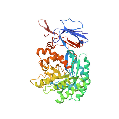
    Cytosine deaminase (CDA) from E. coli is a member of the amidohydrolase superfamily. The structure of the zinc-activated enzyme was determined in the presence of phosphonocytosine, a mimic of the tetrahedral reaction intermediate. This compound inhibits the deamination of cytosine with a K(i) of 52 nM. The zinc- and iron-containing enzymes were characterized to determine the effect of the divalent cations on activation of the hydrolytic water. Fe-CDA loses activity at low pH with a kinetic pK(a) of 6.0, and Zn-CDA has a kinetic pK(a) of 7.3. Mutation of Gln-156 decreased the catalytic activity by more than 5 orders of magnitude, supporting its role in substrate binding. Mutation of Glu-217, Asp-313, and His-246 significantly decreased catalytic activity supporting the role of these three residues in activation of the hydrolytic water molecule and facilitation of proton transfer reactions. A library of potential substrates was used to probe the structural determinants responsible for catalytic activity. CDA was able to catalyze the deamination of isocytosine and the hydrolysis of 3-oxauracil. Large inverse solvent isotope effects were obtained on k(cat) and k(cat)/K(m), consistent with the formation of a low-barrier hydrogen bond during the conversion of cytosine to uracil. A chemical mechanism for substrate deamination by CDA was proposed.


  • Organizational Affiliation
    • Department of Chemistry, Texas A&M University, College Station, Texas 77842-3012, United States.

Macromolecule Content 

  • Total Structure Weight: 49.06 kDa 
  • Atom Count: 3,675 
  • Modeled Residue Count: 422 
  • Deposited Residue Count: 427 
  • Unique protein chains: 1

Macromolecules

Find similar proteins by:|  3D Structure
Entity ID: 1
MoleculeChains  Sequence LengthOrganismDetailsImage
Cytosine deaminase427Escherichia coliMutation(s): 0 
Gene Names: codA
EC: 3.5.4 (UniProt), 3.5.4.1 (UniProt)
UniProt
Find proteins for P25524 (Escherichia coli (strain K12))
Explore P25524 
Go to UniProtKB:  P25524
Entity Groups
Sequence Clusters30% Identity50% Identity70% Identity90% Identity95% Identity100% Identity
UniProt GroupP25524
Sequence Annotations
Expand
Reference Sequence

Small Molecules

Ligands 5 Unique
IDChains Name / Formula / InChI Key2D Diagram3D Interactions
PXN

Query on PXN


Download:Ideal Coordinates CCD File
B [auth A](2S)-1-[3-{[(2R)-2-hydroxypropyl]oxy}-2,2-bis({[(2R)-2-hydroxypropyl]oxy}methyl)propoxy]propan-2-ol
C17 H36 O8
GXEZGLLPFFKHGE-FPCVCCKLSA-N
O7U

Query on O7U


Download:Ideal Coordinates CCD File
E [auth A](2R)-2-amino-2,5-dihydro-1,5,2-diazaphosphinin-6(1H)-one 2-oxide
C3 H6 N3 O2 P
GGLLBAYBJJLFCT-SECBINFHSA-N
PEG

Query on PEG


Download:Ideal Coordinates CCD File
L [auth A],
M [auth A]
DI(HYDROXYETHYL)ETHER
C4 H10 O3
MTHSVFCYNBDYFN-UHFFFAOYSA-N
GOL

Query on GOL


Download:Ideal Coordinates CCD File
F [auth A]
G [auth A]
H [auth A]
I [auth A]
J [auth A]
F [auth A],
G [auth A],
H [auth A],
I [auth A],
J [auth A],
K [auth A]
GLYCEROL
C3 H8 O3
PEDCQBHIVMGVHV-UHFFFAOYSA-N
ZN

Query on ZN


Download:Ideal Coordinates CCD File
C [auth A],
D [auth A]
ZINC ION
Zn
PTFCDOFLOPIGGS-UHFFFAOYSA-N

Experimental Data & Validation

Experimental Data

  • Method: X-RAY DIFFRACTION
  • Resolution: 1.71 Å
  • R-Value Free:  0.193 (Depositor), 0.190 (DCC) 
  • R-Value Work:  0.173 (Depositor), 0.171 (DCC) 
  • R-Value Observed: 0.174 (Depositor) 
Space Group: H 3 2
Unit Cell:
Length ( Å )Angle ( ˚ )
a = 145.212α = 90
b = 145.212β = 90
c = 200.609γ = 120
Software Package:
Software NamePurpose
ADSCdata collection
BALBESphasing
PHENIXrefinement
DENZOdata reduction
SCALEPACKdata scaling

Structure Validation

View Full Validation Report


Ligand Structure Quality Assessment 


Entry History 

Deposition Data

Revision History  (Full details and data files)

  • Version 1.0: 2011-06-15
    Type: Initial release
  • Version 1.1: 2011-07-13
    Changes: Version format compliance
  • Version 1.2: 2011-11-16
    Changes: Atomic model
  • Version 1.3: 2012-03-07
    Changes: Structure summary
  • Version 1.4: 2023-09-06
    Changes: Data collection, Database references, Derived calculations, Refinement description
  • Version 1.5: 2023-10-04
    Changes: Derived calculations