3O6M | pdb_00003o6m

Anti-Tat HIV 11H6H1 Fab' complexed with a 9-mer Tat peptide


Experimental Data Snapshot

  • Method: X-RAY DIFFRACTION
  • Resolution: 2.40 Å
  • R-Value Free: 
    0.290 (Depositor), 0.289 (DCC) 
  • R-Value Work: 
    0.211 (Depositor), 0.211 (DCC) 
  • R-Value Observed: 
    0.215 (Depositor) 

Starting Model: experimental
View more details

wwPDB Validation 3D Report Full Report

Validation slider image for 3O6M

This is version 1.3 of the entry. See complete history

Literature

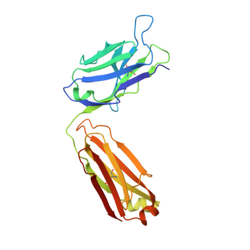
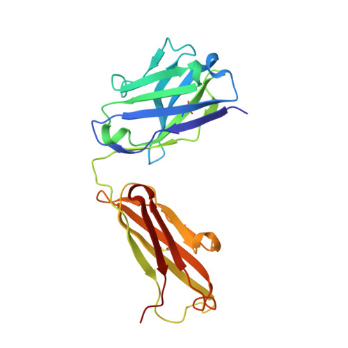
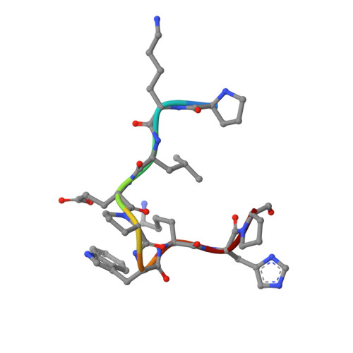
Fab'-induced folding of antigenic N-terminal peptides from intrinsically disordered HIV-1 Tat revealed by X-ray crystallography.

Serriere, J.Dugua, J.M.Bossus, M.Verrier, B.Haser, R.Gouet, P.Guillon, C.

(2011) J Mol Biology 405: 33-42

  • DOI: https://doi.org/10.1016/j.jmb.2010.10.033
  • Primary Citation Related Structures: 
    3O6K, 3O6L, 3O6M

  • PubMed Abstract: 

    Tat, the transcriptional activator protein of human immunodeficiency virus type 1 (HIV-1), is critical for viral replication and is a potential HIV-1 vaccine candidate. This intrinsically disordered protein is present in the extracellular medium and is involved in the pathogenicity of HIV through its interaction with different cellular and viral biological partners. A monoclonal antibody termed 11H6H1, which is specific for the N-terminal region of Tat, was selected for a functional and structural study of the HIV-1 Tat protein. The equilibrium dissociation constants (K(d)) of Tat and Tat fragments complexed with 11H6H1 were estimated by competitive ELISA. Tat contains a single tryptophan residue, Trp11, located in the N-terminal region. We show that the substitution of Trp11 by a phenylalanine completely abolishes the binding of 11H6H1, whereas the transactivating activity of Tat is preserved. The epitope recognized by 11H6H1 was restricted to the 9-mer peptide P(6)KLEPWKHP(14) centered on Trp11. The crystal structures of this 9-mer peptide and of an overlapping 15-mer peptide were determined in complex with Fab' 11H6H1 at 2.4 Å and 2.1 Å resolution, respectively. Tat is intrinsically disordered and can undergo induced folding upon association with a biological partner. Our crystallographic study reveals that the two Tat peptides, which are lodged in the U-shaped groove of the Fab' antigen-binding site, adopt a standard type I β-turn conformation. The central Trp11 that is critical for Fab' recognition is further stabilized by π-stacking interactions. The structural and biological consequences of this induced folding in HIV pathogenesis are discussed.


  • Organizational Affiliation
    • Université de Lyon, IFR128 BioSciences Gerland-Lyon Sud, 7 Passage du Vercors, France.

Macromolecule Content 

  • Total Structure Weight: 48.91 kDa 
  • Atom Count: 3,507 
  • Modeled Residue Count: 443 
  • Deposited Residue Count: 447 
  • Unique protein chains: 3

Macromolecules

Find similar proteins by:|  3D Structure
Entity ID: 1
MoleculeChains  Sequence LengthOrganismDetailsImage
11H6H1 Fab' light chainA [auth L]219Mus musculusMutation(s): 0 
Find similar proteins by:|  3D Structure
Entity ID: 2
MoleculeChains  Sequence LengthOrganismDetailsImage
11H6H1 Fab' heavy chainB [auth H]219Mus musculusMutation(s): 0 
Find similar proteins by:  Sequence   |   3D Structure  
Entity ID: 3
MoleculeChains  Sequence LengthOrganismDetailsImage
Protein Tat 9-mer peptide9N/AMutation(s): 0 
UniProt
Find proteins for Q98XH7 (Human immunodeficiency virus type 1)
Explore Q98XH7 
Go to UniProtKB:  Q98XH7
Entity Groups
UniProt GroupQ98XH7
Sequence Annotations
Expand
Reference Sequence

Experimental Data & Validation

Experimental Data

  • Method: X-RAY DIFFRACTION
  • Resolution: 2.40 Å
  • R-Value Free:  0.290 (Depositor), 0.289 (DCC) 
  • R-Value Work:  0.211 (Depositor), 0.211 (DCC) 
  • R-Value Observed: 0.215 (Depositor) 
Space Group: C 1 2 1
Unit Cell:
Length ( Å )Angle ( ˚ )
a = 123.84α = 90
b = 69.72β = 109.94
c = 62.16γ = 90
Software Package:
Software NamePurpose
ADSCdata collection
AMoREphasing
REFMACrefinement
XDSdata reduction
XSCALEdata scaling

Structure Validation

View Full Validation Report


Entry History 

Deposition Data

Revision History  (Full details and data files)

  • Version 1.0: 2010-11-10
    Type: Initial release
  • Version 1.1: 2011-07-13
    Changes: Version format compliance
  • Version 1.2: 2023-09-06
    Changes: Data collection, Database references, Refinement description
  • Version 1.3: 2024-10-30
    Changes: Structure summary