3O5R

Complex of Fk506 with the Fk1 domain mutant A19T of FKBP51


Experimental Data Snapshot

  • Method: X-RAY DIFFRACTION
  • Resolution: 1.10 Å
  • R-Value Free: 0.157 
  • R-Value Work: 0.123 
  • R-Value Observed: 0.124 

Starting Model: experimental
View more details

wwPDB Validation   3D Report Full Report


Ligand Structure Quality Assessment 


This is version 1.3 of the entry. See complete history


Literature

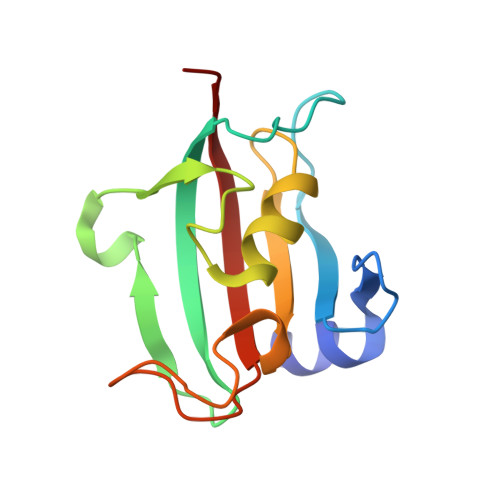
Structural characterization of the PPIase domain of FKBP51, a cochaperone of human Hsp90.

Bracher, A.Kozany, C.Thost, A.K.Hausch, F.

(2011) Acta Crystallogr D Biol Crystallogr 67: 549-559

  • DOI: https://doi.org/10.1107/S0907444911013862
  • Primary Citation of Related Structures:  
    3O5D, 3O5E, 3O5F, 3O5G, 3O5I, 3O5J, 3O5K, 3O5L, 3O5M, 3O5O, 3O5P, 3O5Q, 3O5R

  • PubMed Abstract: 

    Steroid hormone receptors are key components of mammalian stress and sex hormone systems. Many of them rely on the Hsp90 chaperone system for full function and are further fine-tuned by Hsp90-associated peptidyl-prolyl isomerases such as FK506-binding proteins 51 and 52. FK506-binding protein 51 (FKBP51) has been shown to reduce glucocorticoid receptor signalling and has been genetically associated with human stress resilience and with numerous psychiatric disorders. The peptidyl-prolyl isomerase domain of FKBP51 contains a high-affinity binding site for the natural products FK506 and rapamycin and has further been shown to convey most of the inhibitory activity on the glucocorticoid receptor. FKBP51 has therefore become a prime new target for the treatment of stress-related affective disorders that could be amenable to structure-based drug design. Here, a series of high-resolution structures of the peptidyl-prolyl isomerase domain of FKBP51 as well as a cocrystal structure with the prototypic ligand FK506 are described. These structures provide a detailed picture of the drug-binding domain of FKBP51 and the molecular binding mode of its ligand as a starting point for the rational design of improved inhibitors.


  • Organizational Affiliation

    Department of Cellular Biochemistry, Max Planck Institute of Biochemistry, Am Klopferspitz 18, 82152 Martinsried, Germany. bracher@biochem.mpg.de


Macromolecules
Find similar proteins by:  (by identity cutoff)  |  3D Structure
Entity ID: 1
MoleculeChains Sequence LengthOrganismDetailsImage
Peptidyl-prolyl cis-trans isomerase FKBP5128Homo sapiensMutation(s): 0 
Gene Names: AIG6FKBP5FKBP51
EC: 5.2.1.8
UniProt & NIH Common Fund Data Resources
Find proteins for Q13451 (Homo sapiens)
Explore Q13451 
Go to UniProtKB:  Q13451
GTEx:  ENSG00000096060 
Entity Groups  
Sequence Clusters30% Identity50% Identity70% Identity90% Identity95% Identity100% Identity
UniProt GroupQ13451
Sequence Annotations
Expand
  • Reference Sequence
Small Molecules
Ligands 1 Unique
IDChains Name / Formula / InChI Key2D Diagram3D Interactions
FK5
Query on FK5

Download Ideal Coordinates CCD File 
B [auth A]8-DEETHYL-8-[BUT-3-ENYL]-ASCOMYCIN
C44 H69 N O12
QJJXYPPXXYFBGM-LFZNUXCKSA-N
Binding Affinity Annotations 
IDSourceBinding Affinity
FK5 BindingDB:  3O5R Ki: 79 (nM) from 1 assay(s)
Experimental Data & Validation

Experimental Data

  • Method: X-RAY DIFFRACTION
  • Resolution: 1.10 Å
  • R-Value Free: 0.157 
  • R-Value Work: 0.123 
  • R-Value Observed: 0.124 
  • Space Group: P 21 21 21
Unit Cell:
Length ( Å )Angle ( ˚ )
a = 42.051α = 90
b = 54.784β = 90
c = 56.816γ = 90
Software Package:
Software NamePurpose
MOSFLMdata reduction
SCALAdata scaling
SHELXrefinement
PDB_EXTRACTdata extraction
SHELXL-97refinement

Structure Validation

View Full Validation Report


Ligand Structure Quality Assessment 


Entry History 

Deposition Data

Revision History  (Full details and data files)

  • Version 1.0: 2011-06-01
    Type: Initial release
  • Version 1.1: 2011-07-13
    Changes: Version format compliance
  • Version 1.2: 2012-03-28
    Changes: Database references
  • Version 1.3: 2023-09-06
    Changes: Data collection, Database references, Derived calculations, Refinement description