


Crystal Structure of Human Dehydrogenase/Reductase (SDR family) member 4 (DHRS4)
Ugochukwu, E., Bhatia, C., Krojer, T., Vollmar, M., Yue, W.W., Bountra, C., Arrowsmith, C.H., Weigelt, J., Edwards, A., von Delft, F., Oppermann, U.To be published.
Experimental Data Snapshot
Starting Model: experimental
View more details
Entity ID: 1 | |||||
|---|---|---|---|---|---|
| Molecule | Chains | Sequence Length | Organism | Details | Image |
| Dehydrogenase/reductase SDR family member 4 | 261 | Homo sapiens | Mutation(s): 0 Gene Names: DHRS4, UNQ851/PRO1800 EC: 1.1.1.184 | 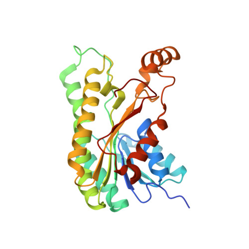 | |
UniProt & NIH Common Fund Data Resources | |||||
GTEx: ENSG00000157326 | |||||
Entity Groups | |||||
| Sequence Clusters | 30% Identity50% Identity70% Identity90% Identity95% Identity100% Identity | ||||
| UniProt Group | Q9BTZ2 | ||||
Sequence AnnotationsExpand | |||||
Reference Sequence | |||||
| Ligands 4 Unique | |||||
|---|---|---|---|---|---|
| ID | Chains | Name / Formula / InChI Key | 2D Diagram | 3D Interactions | |
| NAP Download:Ideal Coordinates CCD File | E [auth A], H [auth B], L [auth C], N [auth D] | NADP NICOTINAMIDE-ADENINE-DINUCLEOTIDE PHOSPHATE C21 H28 N7 O17 P3 XJLXINKUBYWONI-NNYOXOHSSA-N |  | ||
| EPE Download:Ideal Coordinates CCD File | G [auth A] | 4-(2-HYDROXYETHYL)-1-PIPERAZINE ETHANESULFONIC ACID C8 H18 N2 O4 S JKMHFZQWWAIEOD-UHFFFAOYSA-N |  | ||
| GOL Download:Ideal Coordinates CCD File | K [auth B], M [auth C] | GLYCEROL C3 H8 O3 PEDCQBHIVMGVHV-UHFFFAOYSA-N |  | ||
| EDO Download:Ideal Coordinates CCD File | F [auth A], I [auth B], J [auth B], O [auth D] | 1,2-ETHANEDIOL C2 H6 O2 LYCAIKOWRPUZTN-UHFFFAOYSA-N |  | ||
| Length ( Å ) | Angle ( ˚ ) |
|---|---|
| a = 60.07 | α = 90 |
| b = 132.2 | β = 90 |
| c = 133.796 | γ = 90 |
| Software Name | Purpose |
|---|---|
| MAR345 | data collection |
| PHASER | phasing |
| REFMAC | refinement |
| MOSFLM | data reduction |
| SCALA | data scaling |


