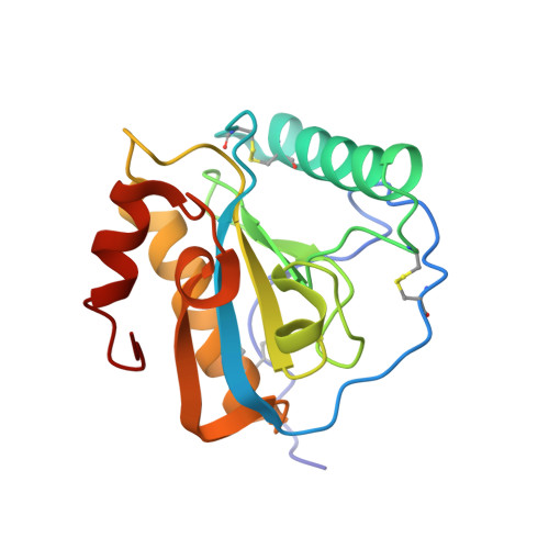
3O4K

Crystal structure of the complex of peptidoglycan recognition protein (PGRP-S) and lipoteichoic acid at 2.1 A resolution


Experimental Data Snapshot

  • Method: X-RAY DIFFRACTION
  • Resolution: 2.11 Å
  • R-Value Free: 0.261 
  • R-Value Work: 0.234 
  • R-Value Observed: 0.234 

Starting Model: experimental
View more details

wwPDB Validation   3D Report Full Report


Ligand Structure Quality Assessment 


This is version 1.4 of the entry. See complete history


Literature

Structural basis of recognition of pathogen-associated molecular patterns and inhibition of proinflammatory cytokines by camel peptidoglycan recognition protein

Sharma, P.Dube, D.Singh, A.Mishra, B.Singh, N.Sinha, M.Dey, S.Kaur, P.Mitra, D.K.Sharma, S.Singh, T.P.

(2011) J Biol Chem 286: 16208-16217

  • DOI: https://doi.org/10.1074/jbc.M111.228163
  • Primary Citation of Related Structures:  
    3O4K, 3RT4

  • PubMed Abstract: 

    Peptidoglycan recognition proteins (PGRPs) are involved in the recognition of pathogen-associated molecular patterns. The well known pathogen-associated molecular patterns include LPS from Gram-negative bacteria and lipoteichoic acid (LTA) from Gram-positive bacteria. In this work, the crystal structures of two complexes of the short form of camel PGRP (CPGRP-S) with LPS and LTA determined at 1.7- and 2.1-Å resolutions, respectively, are reported. Both compounds were held firmly inside the complex formed with four CPGRP-S molecules designated A, B, C, and D. The binding cleft is located at the interface of molecules C and D, which is extendable to the interface of molecules A and C. The interface of molecules A and B is tightly packed, whereas that of molecules B and D forms a wide channel. The hydrophilic moieties of these compounds occupy a common region, whereas hydrophobic chains interact with distinct regions in the binding site. The binding studies showed that CPGRP-S binds to LPS and LTA with affinities of 1.6 × 10(-9) and 2.4 × 10(-8) M, respectively. The flow cytometric studies showed that both LPS- and LTA-induced expression of the proinflammatory cytokines TNF-α and IL-6 was inhibited by CPGRP-S. The results of animal studies using mouse models indicated that both LPS- and LTA-induced mortality rates decreased drastically when CPGRP-S was administered. The recognition of both LPS and LTA, their high binding affinities for CPGRP-S, the significant decrease in the production of LPS- and LTA-induced TNF-α and IL-6, and the drastic reduction in the mortality rates in mice by CPGRP-S indicate its useful properties as an antibiotic agent.


  • Organizational Affiliation

    Department of Biophysics, All India Institute of Medical Sciences, New Delhi, India.


Macromolecules
Find similar proteins by:  (by identity cutoff)  |  3D Structure
Entity ID: 1
MoleculeChains Sequence LengthOrganismDetailsImage
Peptidoglycan recognition protein 1
A, B, C, D
171Camelus dromedariusMutation(s): 0 
UniProt
Find proteins for Q9GK12 (Camelus dromedarius)
Explore Q9GK12 
Go to UniProtKB:  Q9GK12
Entity Groups  
Sequence Clusters30% Identity50% Identity70% Identity90% Identity95% Identity100% Identity
UniProt GroupQ9GK12
Sequence Annotations
Expand
  • Reference Sequence
Small Molecules
Ligands 3 Unique
IDChains Name / Formula / InChI Key2D Diagram3D Interactions
LTC
Query on LTC

Download Ideal Coordinates CCD File 
E [auth C](2S)-1-({3-O-[2-(acetylamino)-4-amino-2,4,6-trideoxy-beta-D-galactopyranosyl]-alpha-D-glucopyranosyl}oxy)-3-(heptanoyloxy)propan-2-yl (7Z)-pentadec-7-enoate
C39 H70 N2 O13
PANDRCFROUDETH-PVQRVOPISA-N
TLA
Query on TLA

Download Ideal Coordinates CCD File 
G [auth D]L(+)-TARTARIC ACID
C4 H6 O6
FEWJPZIEWOKRBE-JCYAYHJZSA-N
GOL
Query on GOL

Download Ideal Coordinates CCD File 
F [auth D]GLYCEROL
C3 H8 O3
PEDCQBHIVMGVHV-UHFFFAOYSA-N
Binding Affinity Annotations 
IDSourceBinding Affinity
LTC PDBBind:  3O4K Kd: 24 (nM) from 1 assay(s)
Experimental Data & Validation

Experimental Data

  • Method: X-RAY DIFFRACTION
  • Resolution: 2.11 Å
  • R-Value Free: 0.261 
  • R-Value Work: 0.234 
  • R-Value Observed: 0.234 
  • Space Group: I 2 2 2
Unit Cell:
Length ( Å )Angle ( ˚ )
a = 88.103α = 90
b = 101.49β = 90
c = 162.8γ = 90
Software Package:
Software NamePurpose
DENZOdata reduction
AMoREphasing
CNSrefinement
AUTOMARdata reduction
SCALEPACKdata scaling

Structure Validation

View Full Validation Report


Ligand Structure Quality Assessment 


Entry History 

Deposition Data

Revision History  (Full details and data files)

  • Version 1.0: 2010-08-25
    Type: Initial release
  • Version 1.1: 2011-07-13
    Changes: Version format compliance
  • Version 1.2: 2011-08-31
    Changes: Database references
  • Version 1.3: 2023-11-01
    Changes: Data collection, Database references, Derived calculations, Refinement description
  • Version 1.4: 2024-11-20
    Changes: Structure summary