3O0F | pdb_00003o0f

Crystal structure of a putative metal-dependent phosphoesterase (BAD_1165) from bifidobacterium adolescentis atcc 15703 at 1.94 A resolution


Experimental Data Snapshot

  • Method: X-RAY DIFFRACTION
  • Resolution: 1.94 Å
  • R-Value Free: 
    0.192 (Depositor), 0.196 (DCC) 
  • R-Value Work: 
    0.160 (Depositor), 0.166 (DCC) 
  • R-Value Observed: 
    0.161 (Depositor) 

wwPDB Validation 3D Report Full Report

Validation slider image for 3O0F

Ligand Structure Quality Assessment 


This is version 1.5 of the entry. See complete history

Literature

Crystal structure of a metal-dependent phosphoesterase (YP_910028.1) from Bifidobacterium adolescentis: Computational prediction and experimental validation of phosphoesterase activity.

Han, G.W.Ko, J.Farr, C.L.Deller, M.C.Xu, Q.Chiu, H.J.Miller, M.D.Sefcikova, J.Somarowthu, S.Beuning, P.J.Elsliger, M.A.Deacon, A.M.Godzik, A.Lesley, S.A.Wilson, I.A.Ondrechen, M.J.

(2011) Proteins 79: 2146-2160

  • DOI: https://doi.org/10.1002/prot.23035
  • Primary Citation Related Structures: 
    3E0F, 3O0F

  • PubMed Abstract: 

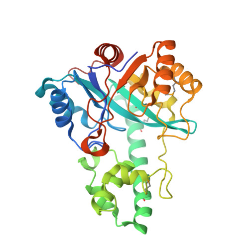
    The crystal structures of an unliganded and adenosine 5'-monophosphate (AMP) bound, metal-dependent phosphoesterase (YP_910028.1) from Bifidobacterium adolescentis are reported at 2.4 and 1.94 Å, respectively. Functional characterization of this enzyme was guided by computational analysis and then confirmed by experiment. The structure consists of a polymerase and histidinol phosphatase (PHP, Pfam: PF02811) domain with a second domain (residues 105-178) inserted in the middle of the PHP sequence. The insert domain functions in binding AMP, but the precise function and substrate specificity of this domain are unknown. Initial bioinformatics analyses yielded multiple potential functional leads, with most of them suggesting DNA polymerase or DNA replication activity. Phylogenetic analysis indicated a potential DNA polymerase function that was somewhat supported by global structural comparisons identifying the closest structural match to the alpha subunit of DNA polymerase III. However, several other functional predictions, including phosphoesterase, could not be excluded. Theoretical microscopic anomalous titration curve shapes, a computational method for the prediction of active sites from protein 3D structures, identified potential reactive residues in YP_910028.1. Further analysis of the predicted active site and local comparison with its closest structure matches strongly suggested phosphoesterase activity, which was confirmed experimentally. Primer extension assays on both normal and mismatched DNA show neither extension nor degradation and provide evidence that YP_910028.1 has neither DNA polymerase activity nor DNA-proofreading activity. These results suggest that many of the sequence neighbors previously annotated as having DNA polymerase activity may actually be misannotated.


  • Organizational Affiliation
    • Joint Center for Structural Genomics, Scripps Research Institute, La Jolla, California 92037, USA.

Macromolecule Content 

  • Total Structure Weight: 33.61 kDa 
  • Atom Count: 2,436 
  • Modeled Residue Count: 287 
  • Deposited Residue Count: 301 
  • Unique protein chains: 1

Macromolecules

Find similar proteins by:|  3D Structure
Entity ID: 1
MoleculeChains  Sequence LengthOrganismDetailsImage
putative metal-dependent phosphoesterase301Bifidobacterium adolescentis ATCC 15703Mutation(s): 0 
Gene Names: BAD_1165
UniProt
Find proteins for A1A2L3 (Bifidobacterium adolescentis (strain ATCC 15703 / DSM 20083 / NCTC 11814 / E194a))
Explore A1A2L3 
Go to UniProtKB:  A1A2L3
Entity Groups
Sequence Clusters30% Identity50% Identity70% Identity90% Identity95% Identity100% Identity
UniProt GroupA1A2L3
Sequence Annotations
Expand
Reference Sequence

Small Molecules

Ligands 5 Unique
IDChains Name / Formula / InChI Key2D Diagram3D Interactions
AMP

Query on AMP


Download:Ideal Coordinates CCD File
B [auth A]ADENOSINE MONOPHOSPHATE
C10 H14 N5 O7 P
UDMBCSSLTHHNCD-KQYNXXCUSA-N
PO4

Query on PO4


Download:Ideal Coordinates CCD File
F [auth A]PHOSPHATE ION
O4 P
NBIIXXVUZAFLBC-UHFFFAOYSA-K
ZN

Query on ZN


Download:Ideal Coordinates CCD File
C [auth A]ZINC ION
Zn
PTFCDOFLOPIGGS-UHFFFAOYSA-N
EDO

Query on EDO


Download:Ideal Coordinates CCD File
G [auth A],
H [auth A]
1,2-ETHANEDIOL
C2 H6 O2
LYCAIKOWRPUZTN-UHFFFAOYSA-N
FE

Query on FE


Download:Ideal Coordinates CCD File
D [auth A],
E [auth A]
FE (III) ION
Fe
VTLYFUHAOXGGBS-UHFFFAOYSA-N
Modified Residues  1 Unique
IDChains TypeFormula2D DiagramParent
MSE
Query on MSE
A
L-PEPTIDE LINKINGC5 H11 N O2 SeMET

Experimental Data & Validation

Experimental Data

  • Method: X-RAY DIFFRACTION
  • Resolution: 1.94 Å
  • R-Value Free:  0.192 (Depositor), 0.196 (DCC) 
  • R-Value Work:  0.160 (Depositor), 0.166 (DCC) 
  • R-Value Observed: 0.161 (Depositor) 
Space Group: P 65
Unit Cell:
Length ( Å )Angle ( ˚ )
a = 103.341α = 90
b = 103.341β = 90
c = 54.744γ = 120
Software Package:
Software NamePurpose
REFMACrefinement
XSCALEdata scaling
PDB_EXTRACTdata extraction
XDSdata reduction

Structure Validation

View Full Validation Report


Ligand Structure Quality Assessment 


Entry History 

Deposition Data

Revision History  (Full details and data files)

  • Version 1.0: 2010-08-25
    Type: Initial release
  • Version 1.1: 2011-07-13
    Changes: Version format compliance
  • Version 1.2: 2011-07-20
    Changes: Database references, Structure summary
  • Version 1.3: 2011-11-16
    Changes: Structure summary
  • Version 1.4: 2023-02-01
    Changes: Database references, Derived calculations
  • Version 1.5: 2024-11-27
    Changes: Data collection, Structure summary