3NZP

Crystal Structure of the Biosynthetic Arginine decarboxylase SpeA from Campylobacter jejuni, Northeast Structural Genomics Consortium Target BR53


Experimental Data Snapshot

  • Method: X-RAY DIFFRACTION
  • Resolution: 3.00 Å
  • R-Value Free: 0.219 
  • R-Value Work: 0.180 
  • R-Value Observed: 0.183 

wwPDB Validation   3D Report Full Report


Ligand Structure Quality Assessment 


This is version 1.2 of the entry. See complete history


Literature

Structures of bacterial biosynthetic arginine decarboxylases.

Forouhar, F.Lew, S.Seetharaman, J.Xiao, R.Acton, T.B.Montelione, G.T.Tong, L.

(2010) Acta Crystallogr Sect F Struct Biol Cryst Commun 66: 1562-1566

  • DOI: https://doi.org/10.1107/S1744309110040649
  • Primary Citation of Related Structures:  
    3NZP, 3NZQ

  • PubMed Abstract: 

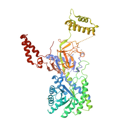
    Biosynthetic arginine decarboxylase (ADC; also known as SpeA) plays an important role in the biosynthesis of polyamines from arginine in bacteria and plants. SpeA is a pyridoxal-5'-phosphate (PLP)-dependent enzyme and shares weak sequence homology with several other PLP-dependent decarboxylases. Here, the crystal structure of PLP-bound SpeA from Campylobacter jejuni is reported at 3.0 Å resolution and that of Escherichia coli SpeA in complex with a sulfate ion is reported at 3.1 Å resolution. The structure of the SpeA monomer contains two large domains, an N-terminal TIM-barrel domain followed by a β-sandwich domain, as well as two smaller helical domains. The TIM-barrel and β-sandwich domains share structural homology with several other PLP-dependent decarboxylases, even though the sequence conservation among these enzymes is less than 25%. A similar tetramer is observed for both C. jejuni and E. coli SpeA, composed of two dimers of tightly associated monomers. The active site of SpeA is located at the interface of this dimer and is formed by residues from the TIM-barrel domain of one monomer and a highly conserved loop in the β-sandwich domain of the other monomer. The PLP cofactor is recognized by hydrogen-bonding, π-stacking and van der Waals interactions.


  • Organizational Affiliation

    Northeast Structural Genomics Consortium, New York, NY 10027, USA.


Macromolecules
Find similar proteins by:  (by identity cutoff)  |  3D Structure
Entity ID: 1
MoleculeChains Sequence LengthOrganismDetailsImage
Arginine decarboxylase
A, B
619Campylobacter jejuni subsp. jejuni NCTC 11168 = ATCC 700819Mutation(s): 0 
Gene Names: Cj0764cspeA
EC: 4.1.1.19
UniProt
Find proteins for Q0PAC6 (Campylobacter jejuni subsp. jejuni serotype O:2 (strain ATCC 700819 / NCTC 11168))
Explore Q0PAC6 
Go to UniProtKB:  Q0PAC6
Entity Groups  
Sequence Clusters30% Identity50% Identity70% Identity90% Identity95% Identity100% Identity
UniProt GroupQ0PAC6
Sequence Annotations
Expand
  • Reference Sequence
Experimental Data & Validation

Experimental Data

  • Method: X-RAY DIFFRACTION
  • Resolution: 3.00 Å
  • R-Value Free: 0.219 
  • R-Value Work: 0.180 
  • R-Value Observed: 0.183 
  • Space Group: P 31 2 1
Unit Cell:
Length ( Å )Angle ( ˚ )
a = 203.071α = 90
b = 203.071β = 90
c = 101.799γ = 120
Software Package:
Software NamePurpose
REFMACrefinement
PDB_EXTRACTdata extraction
ADSCdata collection
HKL-2000data reduction
SCALEPACKdata scaling
SHELXphasing
SOLVEphasing
RESOLVEphasing
CNSrefinement

Structure Validation

View Full Validation Report


Ligand Structure Quality Assessment 


Entry History 

Revision History  (Full details and data files)

  • Version 1.0: 2010-09-01
    Type: Initial release
  • Version 1.1: 2011-07-13
    Changes: Version format compliance
  • Version 1.2: 2012-02-22
    Changes: Structure summary