


Crystal Structure of Phosphoribosylaminoimidazole-Succinocarboxamide Synthase from Clostridium perfringens
Kim, Y., Mulligan, R., Kwon, K., Anderson, W.F., Joachimiak, A.To be published.
Experimental Data Snapshot
Starting Model: experimental
View more details
Entity ID: 1 | |||||
|---|---|---|---|---|---|
| Molecule | Chains | Sequence Length | Organism | Details | Image |
| Phosphoribosylaminoimidazole-succinocarboxamide synthase | 238 | Clostridium perfringens ATCC 13124 | Mutation(s): 0 Gene Names: CPF_0673, purC EC: 6.3.2.6 | 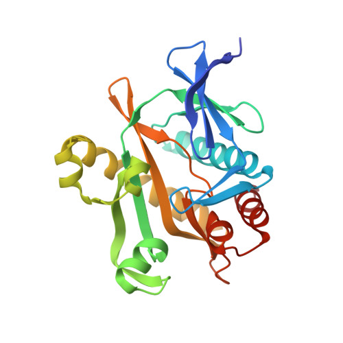 | |
UniProt | |||||
Entity Groups | |||||
| Sequence Clusters | 30% Identity50% Identity70% Identity90% Identity95% Identity100% Identity | ||||
| UniProt Group | Q0TTB4 | ||||
Sequence AnnotationsExpand | |||||
Reference Sequence | |||||
| Ligands 4 Unique | |||||
|---|---|---|---|---|---|
| ID | Chains | Name / Formula / InChI Key | 2D Diagram | 3D Interactions | |
| ADP Download:Ideal Coordinates CCD File | C [auth A], G [auth B], H [auth B] | ADENOSINE-5'-DIPHOSPHATE C10 H15 N5 O10 P2 XTWYTFMLZFPYCI-KQYNXXCUSA-N |  | ||
| AMP Download:Ideal Coordinates CCD File | D [auth A] | ADENOSINE MONOPHOSPHATE C10 H14 N5 O7 P UDMBCSSLTHHNCD-KQYNXXCUSA-N |  | ||
| CIT Download:Ideal Coordinates CCD File | E [auth A], I [auth B] | CITRIC ACID C6 H8 O7 KRKNYBCHXYNGOX-UHFFFAOYSA-N |  | ||
| GOL Download:Ideal Coordinates CCD File | F [auth A] | GLYCEROL C3 H8 O3 PEDCQBHIVMGVHV-UHFFFAOYSA-N |  | ||
| Length ( Å ) | Angle ( ˚ ) |
|---|---|
| a = 80.877 | α = 90 |
| b = 148.913 | β = 90 |
| c = 45.315 | γ = 90 |
| Software Name | Purpose |
|---|---|
| HKL-3000 | data collection |
| HKL-3000 | phasing |
| MOLREP | phasing |
| BALBES | phasing |
| PHENIX | refinement |
| HKL-3000 | data reduction |
| HKL-3000 | data scaling |


