3NR7 | pdb_00003nr7

Crystal structure of S. typhimurium H-NS 1-83


Experimental Data Snapshot

  • Method: X-RAY DIFFRACTION
  • Resolution: 3.70 Å
  • R-Value Free: 
    0.304 (Depositor), 0.302 (DCC) 
  • R-Value Work: 
    0.271 (Depositor), 0.277 (DCC) 
  • R-Value Observed: 
    0.274 (Depositor) 

Starting Model: experimental
View more details

wwPDB Validation 3D Report Full Report

Validation slider image for 3NR7

This is version 1.2 of the entry. See complete history

Literature

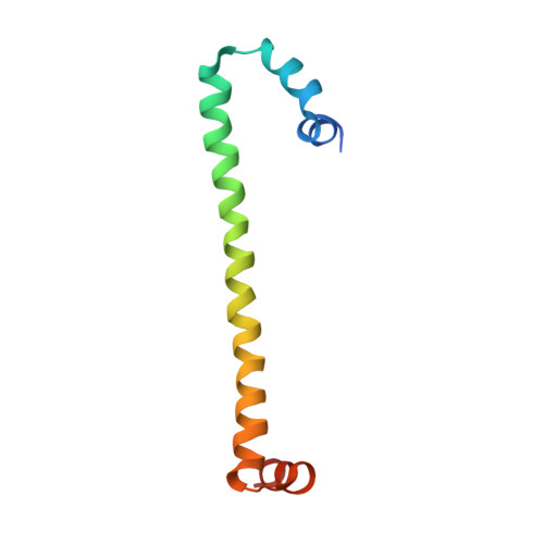
H-NS forms a superhelical protein scaffold for DNA condensation.

Arold, S.T.Leonard, P.G.Parkinson, G.N.Ladbury, J.E.

(2010) Proc Natl Acad Sci U S A 107: 15728-15732

  • DOI: https://doi.org/10.1073/pnas.1006966107
  • Primary Citation Related Structures: 
    3NR7

  • PubMed Abstract: 

    The histone-like nucleoid structuring (H-NS) protein plays a fundamental role in DNA condensation and is a key regulator of enterobacterial gene expression in response to changes in osmolarity, pH, and temperature. The protein is capable of high-order self-association via interactions of its oligomerization domain. Using crystallography, we have solved the structure of this complete domain in an oligomerized state. The observed superhelical structure establishes a mechanism for the self-association of H-NS via both an N-terminal antiparallel coiled-coil and a second, hitherto unidentified, helix-turn-helix dimerization interface at the C-terminal end of the oligomerization domain. The helical scaffold suggests the formation of a H-NS:plectonemic DNA nucleoprotein complex that is capable of explaining published biophysical and functional data, and establishes a unifying structural basis for coordinating the DNA packaging and transcription repression functions of H-NS.


  • Organizational Affiliation
    • Department of Biochemistry and Molecular Biology, University of Texas, M. D. Anderson Cancer Center, Houston, 77030, USA.

Macromolecule Content 

  • Total Structure Weight: 19.59 kDa 
  • Atom Count: 1,288 
  • Modeled Residue Count: 162 
  • Deposited Residue Count: 172 
  • Unique protein chains: 1

Macromolecules

Find similar proteins by:|  3D Structure
Entity ID: 1
MoleculeChains  Sequence LengthOrganismDetailsImage
DNA-binding protein H-NS
A, B
86Salmonella enterica subsp. enterica serovar TyphimuriumMutation(s): 1 
Gene Names: H-NShnshnsAosmZSTM1751
UniProt
Find proteins for P0A1S2 (Salmonella typhimurium (strain LT2 / SGSC1412 / ATCC 700720))
Explore P0A1S2 
Go to UniProtKB:  P0A1S2
Entity Groups
Sequence Clusters30% Identity50% Identity70% Identity90% Identity95% Identity100% Identity
UniProt GroupP0A1S2
Sequence Annotations
Expand
Reference Sequence

Experimental Data & Validation

Experimental Data

  • Method: X-RAY DIFFRACTION
  • Resolution: 3.70 Å
  • R-Value Free:  0.304 (Depositor), 0.302 (DCC) 
  • R-Value Work:  0.271 (Depositor), 0.277 (DCC) 
  • R-Value Observed: 0.274 (Depositor) 
Space Group: P 65
Unit Cell:
Length ( Å )Angle ( ˚ )
a = 130.397α = 90
b = 130.397β = 90
c = 55.061γ = 120
Software Package:
Software NamePurpose
CrystalCleardata collection
MOLREPphasing
REFMACrefinement
d*TREKdata reduction
d*TREKdata scaling

Structure Validation

View Full Validation Report


Entry History 

Deposition Data

Revision History  (Full details and data files)

  • Version 1.0: 2010-09-01
    Type: Initial release
  • Version 1.1: 2011-07-13
    Changes: Version format compliance
  • Version 1.2: 2023-09-06
    Changes: Data collection, Database references, Refinement description