3NOS | pdb_00003nos

HUMAN ENDOTHELIAL NITRIC OXIDE SYNTHASE WITH ARGININE SUBSTRATE


Experimental Data Snapshot

  • Method: X-RAY DIFFRACTION
  • Resolution: 2.40 Å
  • R-Value Free: 
    0.308 (Depositor) 
  • R-Value Work: 
    0.193 (Depositor) 

wwPDB Validation 3D Report Full Report

Validation slider image for 3NOS

This is version 1.3 of the entry. See complete history

Literature

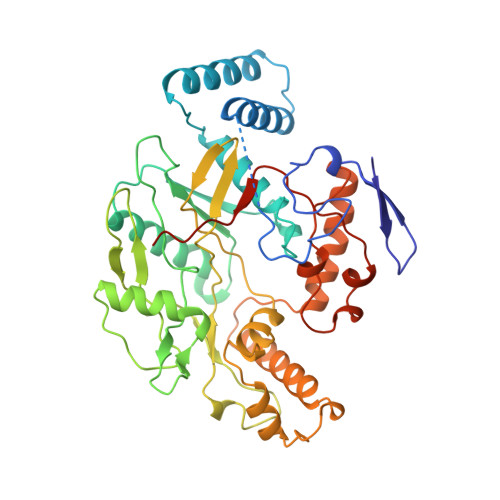
Structural characterization of nitric oxide synthase isoforms reveals striking active-site conservation.

Fischmann, T.O.Hruza, A.Niu, X.D.Fossetta, J.D.Lunn, C.A.Dolphin, E.Prongay, A.J.Reichert, P.Lundell, D.J.Narula, S.K.Weber, P.C.

(1999) Nat Struct Biol 6: 233-242

  • DOI: https://doi.org/10.1038/6675
  • Primary Citation Related Structures: 
    3NOS, 4NOS

  • PubMed Abstract: 

    Crystal structures of human endothelial nitric oxide synthase (eNOS) and human inducible NOS (iNOS) catalytic domains were solved in complex with the arginine substrate and an inhibitor S-ethylisothiourea (SEITU), respectively. The small molecules bind in a narrow cleft within the larger active-site cavity containing heme and tetrahydrobiopterin. Both are hydrogen-bonded to a conserved glutamate (eNOS E361, iNOS E377). The active-site residues of iNOS and eNOS are nearly identical. Nevertheless, structural comparisons provide a basis for design of isozyme-selective inhibitors. The high-resolution, refined structures of eNOS (2.4 A resolution) and iNOS (2.25 A resolution) reveal an unexpected structural zinc situated at the intermolecular interface and coordinated by four cysteines, two from each monomer.


  • Organizational Affiliation
    • Structural Chemistry Department, Schering-Plough Research Institute, Kenilworth, New Jersey 07033, USA. thierry.fischmann@spcorp.com

Macromolecule Content 

  • Total Structure Weight: 98.2 kDa 
  • Atom Count: 7,042 
  • Modeled Residue Count: 800 
  • Deposited Residue Count: 854 
  • Unique protein chains: 1

Macromolecules

Find similar proteins by:|  3D Structure
Entity ID: 1
MoleculeChains  Sequence LengthOrganismDetailsImage
ENDOTHELIAL NITRIC-OXIDE SYNTHASE
A, B
427Homo sapiensMutation(s): 0 
EC: 1.14.13.39
UniProt & NIH Common Fund Data Resources
Find proteins for P29474 (Homo sapiens)
Explore P29474 
Go to UniProtKB:  P29474
PHAROS:  P29474
GTEx:  ENSG00000164867 
Entity Groups
Sequence Clusters30% Identity50% Identity70% Identity90% Identity95% Identity100% Identity
UniProt GroupP29474
Sequence Annotations
Expand
Reference Sequence

Small Molecules

Ligands 4 Unique
IDChains Name / Formula / InChI Key2D Diagram3D Interactions
HEM

Query on HEM


Download:Ideal Coordinates CCD File
E [auth A],
H [auth B]
PROTOPORPHYRIN IX CONTAINING FE
C34 H32 Fe N4 O4
KABFMIBPWCXCRK-RGGAHWMASA-L
H4B

Query on H4B


Download:Ideal Coordinates CCD File
F [auth A],
I [auth B]
5,6,7,8-TETRAHYDROBIOPTERIN
C9 H15 N5 O3
FNKQXYHWGSIFBK-RPDRRWSUSA-N
HAR

Query on HAR


Download:Ideal Coordinates CCD File
D [auth A],
G [auth B]
N-OMEGA-HYDROXY-L-ARGININE
C6 H14 N4 O3
FQWRAVYMZULPNK-BYPYZUCNSA-N
ZN

Query on ZN


Download:Ideal Coordinates CCD File
C [auth A]ZINC ION
Zn
PTFCDOFLOPIGGS-UHFFFAOYSA-N

Experimental Data & Validation

Experimental Data

  • Method: X-RAY DIFFRACTION
  • Resolution: 2.40 Å
  • R-Value Free:  0.308 (Depositor) 
  • R-Value Work:  0.193 (Depositor) 
Space Group: P 21 21 21
Unit Cell:
Length ( Å )Angle ( ˚ )
a = 68.859α = 90
b = 93.264β = 90
c = 156.117γ = 90
Software Package:
Software NamePurpose
AMoREphasing
SHARPphasing
SOLOMONphasing
DMmodel building
X-PLORrefinement
DENZOdata reduction
CCP4data scaling
DMphasing

Structure Validation

View Full Validation Report


Entry History 

Deposition Data

Revision History  (Full details and data files)

  • Version 1.0: 2000-02-04
    Type: Initial release
  • Version 1.1: 2007-10-16
    Changes: Version format compliance
  • Version 1.2: 2011-07-13
    Changes: Derived calculations, Version format compliance
  • Version 1.3: 2023-12-27
    Changes: Data collection, Database references, Derived calculations