Crystal structure and centromere binding of the plasmid segregation protein ParB from pCXC100
Huang, L., Yin, P., Zhu, X., Zhang, Y., Ye, K.(2011) Nucleic Acids Res 39: 2954-2968
- PubMed: 21123191 Search on PubMedSearch on PubMed Central
- DOI: https://doi.org/10.1093/nar/gkq915
- Primary Citation Related Structures:
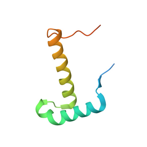
3NO7 - PubMed Abstract:
Plasmid pCXC100 from the Gram-positive bacterium Leifsonia xyli subsp. cynodontis uses a type Ib partition system that includes a centromere region, a Walker-type ATPase ParA and a centromere-binding protein ParB for stable segregation. However, ParB shows no detectable sequence homology to any DNA-binding motif. Here, we study the ParB centromere interaction by structural and biochemical approaches. The crystal structure of the C-terminal DNA-binding domain of ParB at 1.4 Å resolution reveals a dimeric ribbon-helix-helix (RHH) motif, supporting the prevalence of RHH motif in centromere binding. Using hydroxyl radical footprinting and quantitative binding assays, we show that the centromere core comprises nine uninterrupted 9-nt direct repeats that can be successively bound by ParB dimers in a cooperative manner. However, the interaction of ParB with a single subsite requires 18 base pairs covering one immediate repeat as well as two halves of flanking repeats. Through mutagenesis, sequence specificity was determined for each position of an 18-bp subsite. These data suggest an unique centromere recognition mechanism by which the repeat sequence is jointly specified by adjacent ParB dimers bound to an overlapped region.
- State Key Laboratory of Virology, College of Life Sciences, Wuhan University, Wuhan, Hubei 430072, China.
Organizational Affiliation: