


Crystal Structure of HIV-1 Protease in Complex with KNI-10772
Kawasaki, Y., Gabelli, S.B., Amzel, L.M., Freire, E.To be published.
Experimental Data Snapshot
Entity ID: 1 | |||||
|---|---|---|---|---|---|
| Molecule | Chains | Sequence Length | Organism | Details | Image |
| Protease | 99 | Human immunodeficiency virus type 1 (BRU ISOLATE) | Mutation(s): 3 Gene Names: pol EC: 3.4.23.16 | 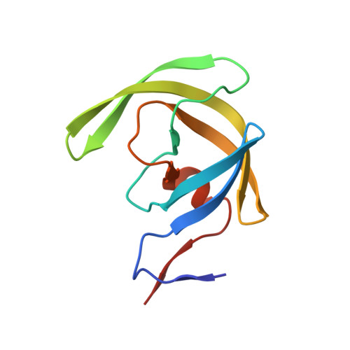 | |
UniProt | |||||
Entity Groups | |||||
| Sequence Clusters | 30% Identity50% Identity70% Identity90% Identity95% Identity100% Identity | ||||
| UniProt Group | P03367 | ||||
Sequence AnnotationsExpand | |||||
Reference Sequence | |||||
| Ligands 4 Unique | |||||
|---|---|---|---|---|---|
| ID | Chains | Name / Formula / InChI Key | 2D Diagram | 3D Interactions | |
| 016 Download:Ideal Coordinates CCD File | E [auth B] | (4R)-3-[(2R,3S)-3-{[(2,6-dimethylphenoxy)acetyl]amino}-2-hydroxy-4-phenylbutanoyl]-N-[(1S,2R)-2-hydroxy-2,3-dihydro-1H-
inden-1-yl]-5,5-dimethyl-1,3-thiazolidine-4-carboxamide C35 H41 N3 O6 S KKTYZYHUPKXLPL-IUHSWKRHSA-N |  | ||
| GOL Download:Ideal Coordinates CCD File | C [auth A] | GLYCEROL C3 H8 O3 PEDCQBHIVMGVHV-UHFFFAOYSA-N |  | ||
| DMS Download:Ideal Coordinates CCD File | F [auth B] | DIMETHYL SULFOXIDE C2 H6 O S IAZDPXIOMUYVGZ-UHFFFAOYSA-N |  | ||
| URE Download:Ideal Coordinates CCD File | D [auth A] | UREA C H4 N2 O XSQUKJJJFZCRTK-UHFFFAOYSA-N |  | ||
| Entity ID: 4 | |||||
|---|---|---|---|---|---|
| ID | Chains | Name | Type/Class | 2D Diagram | 3D Interactions |
| PRD_000572 (016) Query on PRD_000572 | E [auth B] | KNI-10772 | Peptide-like / Inhibitor | | |
| Length ( Å ) | Angle ( ˚ ) |
|---|---|
| a = 57.896 | α = 90 |
| b = 85.389 | β = 90 |
| c = 46.691 | γ = 90 |
| Software Name | Purpose |
|---|---|
| DENZO | data reduction |
| SCALEPACK | data scaling |
| REFMAC | refinement |
| PDB_EXTRACT | data extraction |


