3NLD

Structure of endothelial nitric oxide synthase heme domain complexed with 6-{{(3'S,4'S)-3'-[2"-(3'''-fluorophenethylamino)ethoxy]pyrrolidin-4'-yl}methyl}-4-methylpyridin-2-amine


Experimental Data Snapshot

  • Method: X-RAY DIFFRACTION
  • Resolution: 2.29 Å
  • R-Value Free: 0.292 
  • R-Value Work: 0.222 
  • R-Value Observed: 0.225 

wwPDB Validation   3D Report Full Report


This is version 1.3 of the entry. See complete history


Literature

Exploration of the Active Site of Neuronal Nitric Oxide Synthase by the Design and Synthesis of Pyrrolidinomethyl 2-Aminopyridine Derivatives.

Ji, H.Delker, S.L.Li, H.Martasek, P.Roman, L.J.Poulos, T.L.Silverman, R.B.

(2010) J Med Chem 53: 7804-7824

  • DOI: https://doi.org/10.1021/jm100947x
  • Primary Citation of Related Structures:  
    3NLD, 3NLE, 3NLF, 3NLG, 3NLH, 3NLI, 3NLJ, 3NLK, 3NLM, 3NLN, 3NLO, 3NLP, 3NLQ, 3NLR

  • PubMed Abstract: 

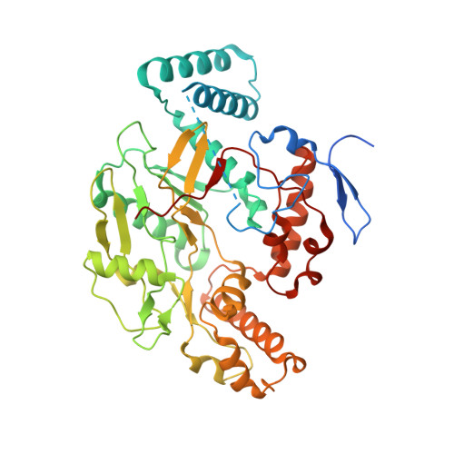
    Neuronal nitric oxide synthase (nNOS) represents an important therapeutic target for the prevention of brain injury and the treatment of various neurodegenerative disorders. A series of trans-substituted amino pyrrolidinomethyl 2-aminopyridine derivatives (8-34) was designed and synthesized. A structure-activity relationship analysis led to the discovery of low nanomolar nNOS inhibitors ((±)-32 and (±)-34) with more than 1000-fold selectivity for nNOS over eNOS. Four enantiomerically pure isomers of 3'-[2''-(3'''-fluorophenethylamino)ethoxy]pyrrolidin-4'-yl}methyl}-4-methylpyridin-2-amine (4) also were synthesized. It was found that (3'R,4'R)-4 can induce enzyme elasticity to generate a new "hot spot" for ligand binding. The inhibitor adopts a unique binding mode, the same as that observed for (3'R,4'R)-3'-[2''-(3'''-fluorophenethylamino)ethylamino]pyrrolidin-4'-yl}methyl}-4-methylpyridin-2-amine ((3'R,4'R)-3) (J. Am. Chem. Soc. 2010, 132 (15), 5437 - 5442). On the basis of structure-activity relationships of 8-34 and different binding conformations of the cis and trans isomers of 3 and 4, critical structural requirements of the NOS active site for ligand binding are revealed.


  • Organizational Affiliation

    Department of Chemistry, Center for Molecular Innovation and Drug Discovery, and Chemistry of Life Processes Institute, Northwestern University, 2145 Sheridan Road, Evanston, Illinois 60208-3113, United States.


Macromolecules
Find similar proteins by:  (by identity cutoff)  |  3D Structure
Entity ID: 1
MoleculeChains Sequence LengthOrganismDetailsImage
Nitric oxide synthase, endothelial
A, B
444Bos taurusMutation(s): 0 
Gene Names: NOS3
EC: 1.14.13.39
UniProt
Find proteins for P29473 (Bos taurus)
Explore P29473 
Go to UniProtKB:  P29473
Entity Groups  
Sequence Clusters30% Identity50% Identity70% Identity90% Identity95% Identity100% Identity
UniProt GroupP29473
Sequence Annotations
Expand
  • Reference Sequence
Small Molecules
Ligands 7 Unique
IDChains Name / Formula / InChI Key2D Diagram3D Interactions
HEM
Query on HEM

Download Ideal Coordinates CCD File 
C [auth A],
J [auth B]
PROTOPORPHYRIN IX CONTAINING FE
C34 H32 Fe N4 O4
KABFMIBPWCXCRK-RGGAHWMASA-L
JSS
Query on JSS

Download Ideal Coordinates CCD File 
G [auth A],
N [auth B]
6-{[(3S,4S)-4-(2-{[2-(3-fluorophenyl)ethyl]amino}ethoxy)pyrrolidin-3-yl]methyl}-4-methylpyridin-2-amine
C21 H29 F N4 O
NESZTSPBRDBHCW-FXAWDEMLSA-N
H4B
Query on H4B

Download Ideal Coordinates CCD File 
D [auth A],
K [auth B]
5,6,7,8-TETRAHYDROBIOPTERIN
C9 H15 N5 O3
FNKQXYHWGSIFBK-RPDRRWSUSA-N
CAD
Query on CAD

Download Ideal Coordinates CCD File 
H [auth A],
O [auth B]
CACODYLIC ACID
C2 H7 As O2
OGGXGZAMXPVRFZ-UHFFFAOYSA-N
GOL
Query on GOL

Download Ideal Coordinates CCD File 
F [auth A],
M [auth B]
GLYCEROL
C3 H8 O3
PEDCQBHIVMGVHV-UHFFFAOYSA-N
ZN
Query on ZN

Download Ideal Coordinates CCD File 
I [auth A]ZINC ION
Zn
PTFCDOFLOPIGGS-UHFFFAOYSA-N
ACT
Query on ACT

Download Ideal Coordinates CCD File 
E [auth A],
L [auth B]
ACETATE ION
C2 H3 O2
QTBSBXVTEAMEQO-UHFFFAOYSA-M
Binding Affinity Annotations 
IDSourceBinding Affinity
JSS BindingDB:  3NLD Ki: min: 3.10e+4, max: 2.62e+7 (nM) from 2 assay(s)
Binding MOAD:  3NLD Ki: 2.62e+4 (nM) from 1 assay(s)
Experimental Data & Validation

Experimental Data

  • Method: X-RAY DIFFRACTION
  • Resolution: 2.29 Å
  • R-Value Free: 0.292 
  • R-Value Work: 0.222 
  • R-Value Observed: 0.225 
  • Space Group: P 21 21 21
Unit Cell:
Length ( Å )Angle ( ˚ )
a = 57.397α = 90
b = 106.885β = 90
c = 157.369γ = 90
Software Package:
Software NamePurpose
REFMACrefinement
CNSrefinement
ADSCdata collection
HKL-2000data reduction
HKL-2000data scaling
CNSphasing

Structure Validation

View Full Validation Report


Entry History 

Deposition Data

Revision History  (Full details and data files)

  • Version 1.0: 2010-11-03
    Type: Initial release
  • Version 1.1: 2011-07-13
    Changes: Version format compliance
  • Version 1.2: 2019-07-17
    Changes: Data collection, Refinement description
  • Version 1.3: 2024-02-21
    Changes: Data collection, Database references, Derived calculations