Molecular adaptability of nucleoside diphosphate kinase b from trypanosomatid parasites: stability, oligomerization and structural determinants of nucleotide binding.
Souza, T.A., Trindade, D.M., Tonoli, C.C., Santos, C.R., Ward, R.J., Arni, R.K., Oliveira, A.H., Murakami, M.T.(2011) Mol Biosyst 7: 2189-2195
- PubMed: 21528129
- DOI: https://doi.org/10.1039/c0mb00307g
- Primary Citation of Related Structures:
3NGR, 3NGS, 3NGT, 3NGU, 3PRV - PubMed Abstract:
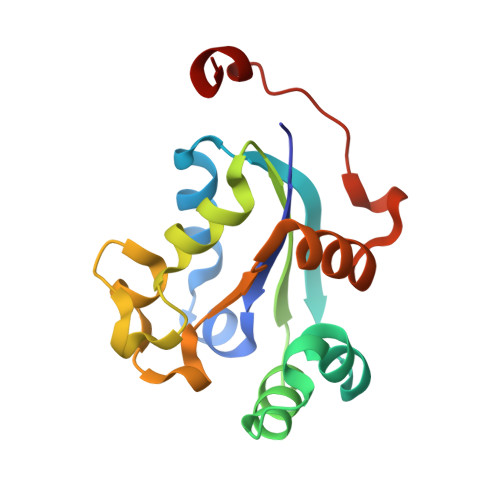
Nucleoside diphosphate kinases play a crucial role in the purine-salvage pathway of trypanosomatid protozoa and have been found in the secretome of Leishmania sp., suggesting a function related to host-cell integrity for the benefit of the parasite. Due to their importance for housekeeping functions in the parasite and by prolonging the life of host cells in infection, they become an attractive target for drug discovery and design. In this work, we describe the first structural characterization of nucleoside diphosphate kinases b from trypanosomatid parasites (tNDKbs) providing insights into their oligomerization, stability and structural determinants for nucleotide binding. Crystallographic studies of LmNDKb when complexed with phosphate, AMP and ADP showed that the crucial hydrogen-bonding residues involved in the nucleotide interaction are fully conserved in tNDKbs. Depending on the nature of the ligand, the nucleotide-binding pocket undergoes conformational changes, which leads to different cavity volumes. SAXS experiments showed that tNDKbs, like other eukaryotic NDKs, form a hexamer in solution and their oligomeric state does not rely on the presence of nucleotides or mimetics. Fluorescence-based thermal-shift assays demonstrated slightly higher stability of tNDKbs compared to human NDKb (HsNDKb), which is in agreement with the fact that tNDKbs are secreted and subjected to variations of temperature in the host cells during infection and disease development. Moreover, tNDKbs were stabilized upon nucleotide binding, whereas HsNDKb was not influenced. Contrasts on the surface electrostatic potential around the nucleotide-binding pocket might be a determinant for nucleotide affinity and protein stability differentiation. All these together demonstrated the molecular adaptation of parasite NDKbs in order to exert their biological functions intra-parasite and when secreted by regulating ATP levels of host cells.
- Laboratório Nacional de Biociências, Laboratório Nacional de Luz Síncrotron, Centro Nacional de Pesquisa em Energia e Materiais, Campinas, SP, Brazil. tatiana.brasil@lnbio.org.br
Organizational Affiliation:


