3NEF | pdb_00003nef

High-resolution pyrabactin-bound PYL1 structure


Experimental Data Snapshot

  • Method: X-RAY DIFFRACTION
  • Resolution: 2.40 Å
  • R-Value Free: 
    0.223 (Depositor), 0.225 (DCC) 
  • R-Value Work: 
    0.188 (Depositor) 
  • R-Value Observed: 
    0.190 (Depositor) 

Starting Model: experimental
View more details

wwPDB Validation 3D Report Full Report

Validation slider image for 3NEF

Ligand Structure Quality Assessment 


This is version 1.2 of the entry. See complete history

Literature

Functional mechanism of the ABA agonist pyrabactin

Hao, Q.Yin, P.Yan, C.Yuan, X.Li, W.Zhang, Z.Liu, L.Wang, J.Yan, N.

(2010) J Biological Chem 285: 28946-28952

  • DOI: https://doi.org/10.1074/jbc.M110.149005
  • Primary Citation Related Structures: 
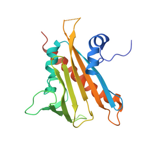
    3NEF, 3NEG

  • PubMed Abstract: 

    Pyrabactin is a synthetic abscisic acid (ABA) agonist that selectively inhibits seed germination. The use of pyrabactin was pivotal in the identification of the PYR1/PYL/RCAR family (PYL) of proteins as the ABA receptor. Although they both act through PYL proteins, pyrabactin and ABA share no apparent chemical or structural similarity. It remains unclear how pyrabactin functions as an ABA agonist. Here, we report the crystal structure of pyrabactin in complex with PYL1 at 2.4 A resolution. Structural and biochemical analyses revealed that recognition of pyrabactin by the pocket residues precedes the closure of switch loop CL2. Structural comparison between pyrabactin- and ABA-bound PYL1 reveals a general principle in the arrangements of function groups of the two distinct ligands. The study provides a framework for the development of novel ABA agonists that may have applicable potentials in agriculture.


  • Organizational Affiliation
    • State Key Laboratory of Biomembrane and Membrane Biotechnology, Tsinghua University, Beijing 100084, China.

Macromolecule Content 

  • Total Structure Weight: 47.03 kDa 
  • Atom Count: 3,015 
  • Modeled Residue Count: 359 
  • Deposited Residue Count: 404 
  • Unique protein chains: 1

Macromolecules

Find similar proteins by:|  3D Structure
Entity ID: 1
MoleculeChains  Sequence LengthOrganismDetailsImage
Abscisic acid receptor PYL1
A, B
202Arabidopsis thalianaMutation(s): 0 
EC: 3.1.3.16
UniProt
Find proteins for Q8VZS8 (Arabidopsis thaliana)
Explore Q8VZS8 
Go to UniProtKB:  Q8VZS8
Entity Groups
Sequence Clusters30% Identity50% Identity70% Identity90% Identity95% Identity100% Identity
UniProt GroupQ8VZS8
Sequence Annotations
Expand
Reference Sequence

Small Molecules

Ligands 1 Unique
IDChains Name / Formula / InChI Key2D Diagram3D Interactions
PYV

Query on PYV


Download:Ideal Coordinates CCD File
C [auth A]4-bromo-N-(pyridin-2-ylmethyl)naphthalene-1-sulfonamide
C16 H13 Br N2 O2 S
GJSDYQXOSHKOGX-UHFFFAOYSA-N

Experimental Data & Validation

Experimental Data

  • Method: X-RAY DIFFRACTION
  • Resolution: 2.40 Å
  • R-Value Free:  0.223 (Depositor), 0.225 (DCC) 
  • R-Value Work:  0.188 (Depositor) 
  • R-Value Observed: 0.190 (Depositor) 
Space Group: P 31 2 1
Unit Cell:
Length ( Å )Angle ( ˚ )
a = 128.57α = 90
b = 128.57β = 90
c = 87.24γ = 120
Software Package:
Software NamePurpose
BSSdata collection
PHASERphasing
PHENIXrefinement
MOSFLMdata reduction
SCALAdata scaling

Structure Validation

View Full Validation Report


Ligand Structure Quality Assessment 


Entry History 

Deposition Data

  • Released Date: 2010-06-16 
  • Deposition Author(s): Yan, N.

Revision History  (Full details and data files)

  • Version 1.0: 2010-06-16
    Type: Initial release
  • Version 1.1: 2011-07-13
    Changes: Version format compliance
  • Version 1.2: 2023-11-01
    Changes: Data collection, Database references, Derived calculations, Refinement description