3NED | pdb_00003ned

mRouge


Experimental Data Snapshot

  • Method: X-RAY DIFFRACTION
  • Resolution: 0.95 Å
  • R-Value Free: 
    0.157 (Depositor), 0.159 (DCC) 
  • R-Value Work: 
    0.133 (Depositor), 0.135 (DCC) 
  • R-Value Observed: 
    0.135 (Depositor) 

wwPDB Validation 3D Report Full Report

Validation slider image for 3NED

This is version 3.0 of the entry. See complete history

Literature

Generation of longer emission wavelength red fluorescent proteins using computationally designed libraries.

Chica, R.A.Moore, M.M.Allen, B.D.Mayo, S.L.

(2010) Proc Natl Acad Sci U S A 107: 20257-20262

  • DOI: https://doi.org/10.1073/pnas.1013910107
  • Primary Citation Related Structures: 
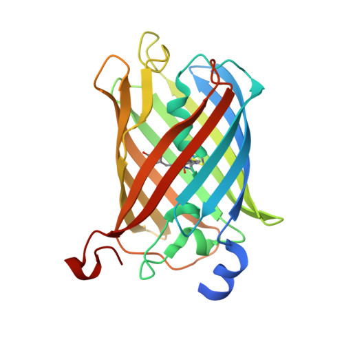
    3NED, 3NEZ

  • PubMed Abstract: 

    The longer emission wavelengths of red fluorescent proteins (RFPs) make them attractive for whole-animal imaging because cells are more transparent to red light. Although several useful RFPs have been developed using directed evolution, the quest for further red-shifted and improved RFPs continues. Herein, we report a structure-based rational design approach to red-shift the fluorescence emission of RFPs. We applied a combined computational and experimental approach that uses computational protein design as an in silico prescreen to generate focused combinatorial libraries of mCherry mutants. The computational procedure helped us identify residues that could fulfill interactions hypothesized to cause red-shifts without destabilizing the protein fold. These interactions include stabilization of the excited state through H-bonding to the acylimine oxygen atom, destabilization of the ground state by hydrophobic packing around the charged phenolate, and stabilization of the excited state by a π-stacking interaction. Our methodology allowed us to identify three mCherry mutants (mRojoA, mRojoB, and mRouge) that display emission wavelengths > 630 nm, representing red-shifts of 20-26 nm. Moreover, our approach required the experimental screening of a total of ∼5,000 clones, a number several orders of magnitude smaller than those previously used to achieve comparable red-shifts. Additionally, crystal structures of mRojoA and mRouge allowed us to verify fulfillment of the interactions hypothesized to cause red-shifts, supporting their contribution to the observed red-shifts.


  • Organizational Affiliation
    • Division of Biology, California Institute of Technology, 1200 East California Boulevard, Pasadena, CA 91125, USA.

Macromolecule Content 

  • Total Structure Weight: 27.88 kDa 
  • Atom Count: 2,617 
  • Modeled Residue Count: 231 
  • Deposited Residue Count: 244 
  • Unique protein chains: 1

Macromolecules

Find similar proteins by:|  3D Structure
Entity ID: 1
MoleculeChains  Sequence LengthOrganismDetailsImage
PAmCherry1 protein242Discosoma sp.Mutation(s): 14 
Gene Names: PAmCherryPAmCherry1
UniProt
Find proteins for D1MPT3 (Discosoma sp.)
Explore D1MPT3 
Go to UniProtKB:  D1MPT3
Entity Groups
Sequence Clusters30% Identity50% Identity70% Identity90% Identity95% Identity100% Identity
UniProt GroupD1MPT3
Sequence Annotations
Expand
Reference Sequence

Experimental Data & Validation

Experimental Data

  • Method: X-RAY DIFFRACTION
  • Resolution: 0.95 Å
  • R-Value Free:  0.157 (Depositor), 0.159 (DCC) 
  • R-Value Work:  0.133 (Depositor), 0.135 (DCC) 
  • R-Value Observed: 0.135 (Depositor) 
Space Group: P 1 21 1
Unit Cell:
Length ( Å )Angle ( ˚ )
a = 48.807α = 90
b = 42.76β = 112.35
c = 61.203γ = 90
Software Package:
Software NamePurpose
iMOSFLMdata reduction
REFMACrefinement
ACORNphasing

Structure Validation

View Full Validation Report


Entry History 

Deposition Data

Revision History  (Full details and data files)

  • Version 1.0: 2010-11-24
    Type: Initial release
  • Version 1.1: 2011-07-13
    Changes: Version format compliance
  • Version 1.2: 2014-01-08
    Changes: Database references
  • Version 1.3: 2015-03-04
    Changes: Derived calculations
  • Version 1.4: 2017-11-08
    Changes: Advisory, Refinement description
  • Version 1.5: 2017-11-15
    Changes: Database references
  • Version 2.0: 2023-11-15
    Changes: Advisory, Atomic model, Data collection, Database references, Derived calculations
  • Version 3.0: 2026-03-18
    Changes: Polymer sequence, Structure summary