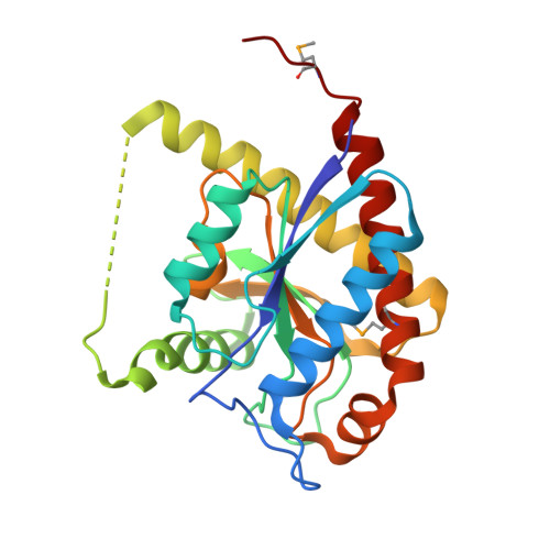
3NE8

The crystal structure of a domain from N-acetylmuramoyl-l-alanine amidase of Bartonella henselae str. Houston-1


Experimental Data Snapshot

  • Method: X-RAY DIFFRACTION
  • Resolution: 1.24 Å
  • R-Value Free: 0.174 
  • R-Value Work: 0.161 
  • R-Value Observed: 0.161 

wwPDB Validation   3D Report Full Report


This is version 1.3 of the entry. See complete history


Literature

A conformational switch controls cell wall-remodelling enzymes required for bacterial cell division.

Yang, D.C.Tan, K.Joachimiak, A.Bernhardt, T.G.

(2012) Mol Microbiol 85: 768-781

  • DOI: https://doi.org/10.1111/j.1365-2958.2012.08138.x
  • Primary Citation of Related Structures:  
    3NE8

  • PubMed Abstract: 

    Remodelling of the peptidoglycan (PG) exoskeleton is intimately tied to the growth and division of bacteria. Enzymes that hydrolyse PG are critical for these processes, but their activities must be tightly regulated to prevent the generation of lethal breaches in the PG matrix. Despite their importance, the mechanisms regulating PG hydrolase activity have remained elusive. Here we investigate the control of cell division hydrolases called amidases (AmiA, AmiB and AmiC) required for Escherichia coli cell division. Poorly regulated amiB mutants were isolated encoding lytic AmiB variants with elevated basal PG hydrolase activities in vitro. The structure of an AmiB orthologue was also solved, revealing that the active site of AmiB is occluded by a conserved alpha helix. Strikingly, most of the amino acid substitutions in the lytic AmiB variants mapped to this domain and are predicted to disrupt its interaction with the active site. Our results therefore support a model in which cell separation is stimulated by the reversible relief of amidase autoinhibition governed by conserved subcomplexes within the cytokinetic ring. Analogous conformational control mechanisms are likely to be part of a general strategy used to control PG hydrolases present within multienzyme PG-remodelling machines.


  • Organizational Affiliation

    Department of Microbiology and Immunobiology, Harvard Medical School, Boston, MA 02115, USA.


Macromolecules
Find similar proteins by:  (by identity cutoff)  |  3D Structure
Entity ID: 1
MoleculeChains Sequence LengthOrganismDetailsImage
N-acetylmuramoyl-l-alanine amidase234Bartonella henselaeMutation(s): 0 
Gene Names: amiBBH08710
EC: 3.5.1.28
UniProt
Find proteins for A0A0H3M3F4 (Bartonella henselae (strain ATCC 49882 / DSM 28221 / CCUG 30454 / Houston 1))
Explore A0A0H3M3F4 
Go to UniProtKB:  A0A0H3M3F4
Entity Groups  
Sequence Clusters30% Identity50% Identity70% Identity90% Identity95% Identity100% Identity
UniProt GroupA0A0H3M3F4
Sequence Annotations
Expand
  • Reference Sequence
Small Molecules
Ligands 4 Unique
IDChains Name / Formula / InChI Key2D Diagram3D Interactions
GOL
Query on GOL

Download Ideal Coordinates CCD File 
G [auth A],
H [auth A],
I [auth A]
GLYCEROL
C3 H8 O3
PEDCQBHIVMGVHV-UHFFFAOYSA-N
ZN
Query on ZN

Download Ideal Coordinates CCD File 
B [auth A]ZINC ION
Zn
PTFCDOFLOPIGGS-UHFFFAOYSA-N
ACT
Query on ACT

Download Ideal Coordinates CCD File 
C [auth A],
D [auth A],
E [auth A]
ACETATE ION
C2 H3 O2
QTBSBXVTEAMEQO-UHFFFAOYSA-M
FMT
Query on FMT

Download Ideal Coordinates CCD File 
F [auth A]FORMIC ACID
C H2 O2
BDAGIHXWWSANSR-UHFFFAOYSA-N
Modified Residues  1 Unique
IDChains TypeFormula2D DiagramParent
MSE
Query on MSE
A
L-PEPTIDE LINKINGC5 H11 N O2 SeMET
Experimental Data & Validation

Experimental Data

  • Method: X-RAY DIFFRACTION
  • Resolution: 1.24 Å
  • R-Value Free: 0.174 
  • R-Value Work: 0.161 
  • R-Value Observed: 0.161 
  • Space Group: C 1 2 1
Unit Cell:
Length ( Å )Angle ( ˚ )
a = 71.185α = 90
b = 53.511β = 90.46
c = 58.495γ = 90
Software Package:
Software NamePurpose
SBC-Collectdata collection
SHELXDphasing
MLPHAREphasing
DMmodel building
ARPmodel building
WARPmodel building
HKL-3000phasing
PHENIXrefinement
HKL-3000data reduction
HKL-3000data scaling
DMphasing

Structure Validation

View Full Validation Report


Entry History 

Deposition Data

Revision History  (Full details and data files)

  • Version 1.0: 2010-07-14
    Type: Initial release
  • Version 1.1: 2011-07-13
    Changes: Version format compliance
  • Version 1.2: 2012-11-07
    Changes: Database references
  • Version 1.3: 2024-11-06
    Changes: Data collection, Database references, Derived calculations, Structure summary