3NE0 | pdb_00003ne0

Structure and functional regulation of RipA, a mycobacterial enzyme essential for daughter cell separation


Experimental Data Snapshot

  • Method: X-RAY DIFFRACTION
  • Resolution: 1.00 Å
  • R-Value Free: 
    0.137 (Depositor), 0.140 (DCC) 
  • R-Value Work: 
    0.104 (Depositor), 0.113 (DCC) 
  • R-Value Observed: 
    0.105 (Depositor) 

wwPDB Validation 3D Report Full Report

Validation slider image for 3NE0

This is version 1.2 of the entry. See complete history

Literature

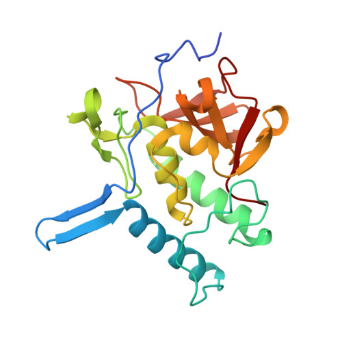
Structure and Functional Regulation of RipA, a Mycobacterial Enzyme Essential for Daughter Cell Separation.

Ruggiero, A.Marasco, D.Squeglia, F.Soldini, S.Pedone, E.Pedone, C.Berisio, R.

(2010) Structure 18: 1184-1190

  • DOI: https://doi.org/10.1016/j.str.2010.06.007
  • Primary Citation Related Structures: 
    3NE0

  • PubMed Abstract: 

    Cell separation depends on cell-wall hydrolases that cleave the peptidoglycan layer connecting daughter cells. In Mycobacterium tuberculosis, this process is governed by the predicted endopeptidase RipA. In the absence of this enzyme, the bacterium is unable to divide and exhibits an abnormal phenotype. We here report the crystal structure of a relevant portion of RipA, containing its catalytic-domain and an extra-domain of hitherto unknown function. The structure clearly demonstrates that RipA is produced as a zymogen, which needs to be activated to achieve cell-division. Bacterial cell-wall degradation assays and proteolysis experiments strongly suggest that activation occurs via proteolytic processing of a fully solvent exposed loop identified in the crystal structure. Indeed, proteolytic cleavage at this loop produces an activated form, consisting of the sole catalytic domain. Our work provides the first evidence of self-inhibition in cell-disconnecting enzymes and opens a field for the design of novel antitubercular therapeutics.


  • Organizational Affiliation
    • Institute of Biostructures and Bioimaging, CNR, Via Mezzocannone 16, I-80134, Naples, Italy.

Macromolecule Content 

  • Total Structure Weight: 22.55 kDa 
  • Atom Count: 1,941 
  • Modeled Residue Count: 208 
  • Deposited Residue Count: 214 
  • Unique protein chains: 1

Macromolecules

Find similar proteins by:|  3D Structure
Entity ID: 1
MoleculeChains  Sequence LengthOrganismDetailsImage
Resuscitation promoting factor Interacting Protein A214Mycobacterium tuberculosis H37RvMutation(s): 0 
Gene Names: rv1477
EC: 3.4
UniProt
Find proteins for O53168 (Mycobacterium tuberculosis (strain ATCC 25618 / H37Rv))
Explore O53168 
Go to UniProtKB:  O53168
Entity Groups
Sequence Clusters30% Identity50% Identity70% Identity90% Identity95% Identity100% Identity
UniProt GroupO53168
Sequence Annotations
Expand
Reference Sequence

Experimental Data & Validation

Experimental Data

  • Method: X-RAY DIFFRACTION
  • Resolution: 1.00 Å
  • R-Value Free:  0.137 (Depositor), 0.140 (DCC) 
  • R-Value Work:  0.104 (Depositor), 0.113 (DCC) 
  • R-Value Observed: 0.105 (Depositor) 
Space Group: P 21 21 21
Unit Cell:
Length ( Å )Angle ( ˚ )
a = 36.866α = 90
b = 65.56β = 90
c = 67.931γ = 90
Software Package:
Software NamePurpose
HKL-2000data collection
SHELXmodel building
SHELXL-97refinement
HKL-2000data reduction
HKL-2000data scaling
SHELXphasing

Structure Validation

View Full Validation Report


Entry History 

Deposition Data

Revision History  (Full details and data files)

  • Version 1.0: 2010-09-22
    Type: Initial release
  • Version 1.1: 2011-07-13
    Changes: Version format compliance
  • Version 1.2: 2024-02-21
    Changes: Data collection, Database references