3NCU | pdb_00003ncu

Structural and functional insights into pattern recognition by the innate immune receptor RIG-I


Experimental Data Snapshot

  • Method: X-RAY DIFFRACTION
  • Resolution: 2.55 Å
  • R-Value Free: 
    0.229 (Depositor), 0.225 (DCC) 
  • R-Value Work: 
    0.190 (Depositor), 0.186 (DCC) 
  • R-Value Observed: 
    0.192 (Depositor) 

Starting Model: experimental
View more details

wwPDB Validation 3D Report Full Report

Validation slider image for 3NCU

This is version 1.2 of the entry. See complete history

Literature

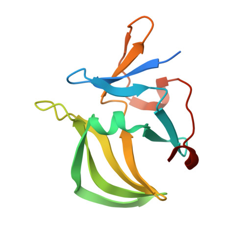
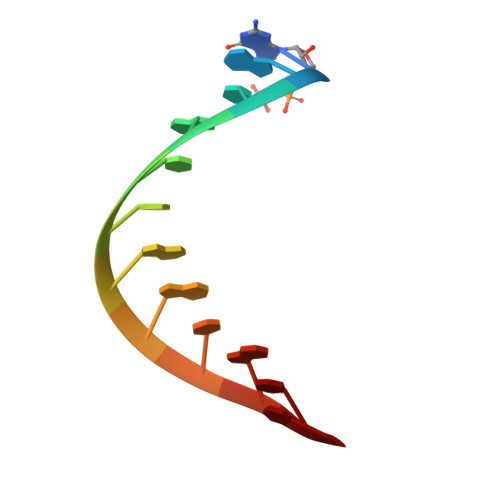
Structural and functional insights into 5'-ppp RNA pattern recognition by the innate immune receptor RIG-I.

Wang, Y.Ludwig, J.Schuberth, C.Goldeck, M.Schlee, M.Li, H.Juranek, S.Sheng, G.Micura, R.Tuschl, T.Hartmann, G.Patel, D.J.

(2010) Nat Struct Mol Biol 17: 781-787

  • DOI: https://doi.org/10.1038/nsmb.1863
  • Primary Citation Related Structures: 
    3NCU

  • PubMed Abstract: 

    RIG-I is a cytosolic helicase that senses 5'-ppp RNA contained in negative-strand RNA viruses and triggers innate antiviral immune responses. Calorimetric binding studies established that the RIG-I C-terminal regulatory domain (CTD) binds to blunt-end double-stranded 5'-ppp RNA a factor of 17 more tightly than to its single-stranded counterpart. Here we report on the crystal structure of RIG-I CTD bound to both blunt ends of a self-complementary 5'-ppp dsRNA 12-mer, with interactions involving 5'-pp clearly visible in the complex. The structure, supported by mutation studies, defines how a lysine-rich basic cleft within the RIG-I CTD sequesters the observable 5'-pp of the bound RNA, with a stacked phenylalanine capping the terminal base pair. Key intermolecular interactions observed in the crystalline state are retained in the complex of 5'-ppp dsRNA 24-mer and full-length RIG-I under in vivo conditions, as evaluated from the impact of binding pocket RIG-I mutations and 2'-OCH(3) RNA modifications on the interferon response.


  • Organizational Affiliation
    • Structural Biology Program, Memorial Sloan-Kettering Cancer Center, New York, New York, USA.

Macromolecule Content 

  • Total Structure Weight: 39.26 kDa 
  • Atom Count: 2,604 
  • Modeled Residue Count: 264 
  • Deposited Residue Count: 292 
  • Unique protein chains: 1
  • Unique nucleic acid chains: 1

Macromolecules


Find similar proteins by:|  3D Structure
Entity ID: 1
MoleculeChains  Sequence LengthOrganismDetailsImage
RIG-I
A, B
134Homo sapiensMutation(s): 0 
Gene Names: DDX58RIG-I
EC: 3.6.1 (PDB Primary Data), 3.6.4.13 (UniProt)
UniProt & NIH Common Fund Data Resources
Find proteins for O95786 (Homo sapiens)
Explore O95786 
Go to UniProtKB:  O95786
PHAROS:  O95786
GTEx:  ENSG00000107201 
Entity Groups
Sequence Clusters30% Identity50% Identity70% Identity90% Identity95% Identity100% Identity
UniProt GroupO95786
Sequence Annotations
Expand
Reference Sequence
Find similar nucleic acids by:  Sequence
Entity ID: 2
MoleculeChains LengthOrganismImage
5'-R(*(GDP)P*AP*CP*GP*CP*UP*AP*GP*CP*GP*UP*C)-3'
C, D
12N/A
Sequence Annotations
Expand
Reference Sequence

Experimental Data & Validation

Experimental Data

  • Method: X-RAY DIFFRACTION
  • Resolution: 2.55 Å
  • R-Value Free:  0.229 (Depositor), 0.225 (DCC) 
  • R-Value Work:  0.190 (Depositor), 0.186 (DCC) 
  • R-Value Observed: 0.192 (Depositor) 
Space Group: P 65
Unit Cell:
Length ( Å )Angle ( ˚ )
a = 83.462α = 90
b = 83.462β = 90
c = 110.364γ = 120
Software Package:
Software NamePurpose
HKL-2000data collection
PHASERphasing
PHENIXrefinement
HKL-2000data reduction
HKL-2000data scaling

Structure Validation

View Full Validation Report


Entry History 

Deposition Data

Revision History  (Full details and data files)

  • Version 1.0: 2010-06-30
    Type: Initial release
  • Version 1.1: 2011-07-13
    Changes: Version format compliance
  • Version 1.2: 2023-09-06
    Changes: Data collection, Database references, Derived calculations, Refinement description