


Crystal structure of S-adenosyl-L-homocysteine hydrolase from brucella melitensis in ternary complex with NAD and adenosine, orthorhombic form
SSGCID, Gardberg, A., Sankaran, B., Abendroth, J., Staker, B.To be published.
Experimental Data Snapshot
Starting Model: experimental
View more details
Entity ID: 1 | |||||
|---|---|---|---|---|---|
| Molecule | Chains | Sequence Length | Organism | Details | Image |
| Adenosylhomocysteinase | 464 | Brucella abortus 2308 | Mutation(s): 0 Gene Names: ahcY, BAB1_2099 EC: 3.3.1.1 (PDB Primary Data), 3.13.2.1 (UniProt) | 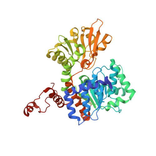 | |
UniProt | |||||
Entity Groups | |||||
| Sequence Clusters | 30% Identity50% Identity70% Identity90% Identity95% Identity100% Identity | ||||
| UniProt Group | Q2YQX8 | ||||
Sequence AnnotationsExpand | |||||
Reference Sequence | |||||
| Ligands 3 Unique | |||||
|---|---|---|---|---|---|
| ID | Chains | Name / Formula / InChI Key | 2D Diagram | 3D Interactions | |
| NAD Download:Ideal Coordinates CCD File | G [auth A], H [auth B], K [auth C], N [auth D] | NICOTINAMIDE-ADENINE-DINUCLEOTIDE C21 H27 N7 O14 P2 BAWFJGJZGIEFAR-NNYOXOHSSA-N |  | ||
| ADN Download:Ideal Coordinates CCD File | F [auth A], J [auth C], M [auth D] | ADENOSINE C10 H13 N5 O4 OIRDTQYFTABQOQ-KQYNXXCUSA-N |  | ||
| K Download:Ideal Coordinates CCD File | E [auth A], I [auth C], L [auth D] | POTASSIUM ION K NPYPAHLBTDXSSS-UHFFFAOYSA-N |  | ||
| Length ( Å ) | Angle ( ˚ ) |
|---|---|
| a = 68.45 | α = 90 |
| b = 165.23 | β = 90 |
| c = 184.14 | γ = 90 |
| Software Name | Purpose |
|---|---|
| ADSC | data collection |
| PHASER | phasing |
| PHENIX | refinement |
| XDS | data reduction |
| XSCALE | data scaling |


