3MWD

Truncated Human ATP-Citrate Lyase with Citrate Bound


Experimental Data Snapshot

  • Method: X-RAY DIFFRACTION
  • Resolution: 2.10 Å
  • R-Value Free: 0.229 
  • R-Value Work: 0.191 
  • R-Value Observed: 0.194 

wwPDB Validation   3D Report Full Report


This is version 1.1 of the entry. See complete history


Literature

Identification of the citrate-binding site of human ATP-citrate lyase using X-ray crystallography.

Sun, T.Hayakawa, K.Bateman, K.S.Fraser, M.E.

(2010) J Biol Chem 285: 27418-27428

  • DOI: https://doi.org/10.1074/jbc.M109.078667
  • Primary Citation of Related Structures:  
    3MWD, 3MWE

  • PubMed Abstract: 

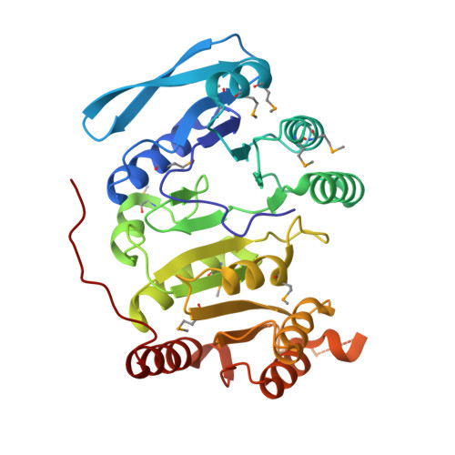
    ATP-citrate lyase (ACLY) catalyzes the conversion of citrate and CoA into acetyl-CoA and oxaloacetate, coupled with the hydrolysis of ATP. In humans, ACLY is the cytoplasmic enzyme linking energy metabolism from carbohydrates to the production of fatty acids. In situ proteolysis of full-length human ACLY gave crystals of a truncated form, revealing the conformations of residues 2-425, 487-750, and 767-820 of the 1101-amino acid protein. Residues 2-425 form three domains homologous to the beta-subunit of succinyl-CoA synthetase (SCS), while residues 487-820 form two domains homologous to the alpha-subunit of SCS. The crystals were grown in the presence of tartrate or the substrate, citrate, and the structure revealed the citrate-binding site. A loop formed by residues 343-348 interacts via specific hydrogen bonds with the hydroxyl and carboxyl groups on the prochiral center of citrate. Arg-379 forms a salt bridge with the pro-R carboxylate of citrate. The pro-S carboxylate is free to react, providing insight into the stereospecificity of ACLY. Because this is the first structure of any member of the acyl-CoA synthetase (NDP-forming) superfamily in complex with its organic acid substrate, locating the citrate-binding site is significant for understanding the catalytic mechanism of each member, including the prototype SCS. Comparison of the CoA-binding site of SCSs with the similar structure in ACLY showed that ACLY possesses a different CoA-binding site. Comparisons of the nucleotide-binding site of SCSs with the similar structure in ACLY indicates that this is the ATP-binding site of ACLY.


  • Organizational Affiliation

    Department of Biological Sciences, University of Calgary, Calgary, Alberta T2N 1N4, Canada.


Macromolecules
Find similar proteins by:  (by identity cutoff)  |  3D Structure
Entity ID: 1
MoleculeChains Sequence LengthOrganismDetailsImage
ATP-citrate synthase425Homo sapiensMutation(s): 0 
Gene Names: ACLY
EC: 2.3.3.8
UniProt & NIH Common Fund Data Resources
Find proteins for P53396 (Homo sapiens)
Explore P53396 
Go to UniProtKB:  P53396
PHAROS:  P53396
GTEx:  ENSG00000131473 
Entity Groups  
Sequence Clusters30% Identity50% Identity70% Identity90% Identity95% Identity100% Identity
UniProt GroupP53396
Sequence Annotations
Expand
  • Reference Sequence
Find similar proteins by:  (by identity cutoff)  |  3D Structure
Entity ID: 2
MoleculeChains Sequence LengthOrganismDetailsImage
ATP-citrate synthase334Homo sapiensMutation(s): 0 
Gene Names: ACLY
EC: 2.3.3.8
UniProt & NIH Common Fund Data Resources
Find proteins for P53396 (Homo sapiens)
Explore P53396 
Go to UniProtKB:  P53396
PHAROS:  P53396
GTEx:  ENSG00000131473 
Entity Groups  
Sequence Clusters30% Identity50% Identity70% Identity90% Identity95% Identity100% Identity
UniProt GroupP53396
Sequence Annotations
Expand
  • Reference Sequence
Small Molecules
Ligands 1 Unique
IDChains Name / Formula / InChI Key2D Diagram3D Interactions
CIT
Query on CIT

Download Ideal Coordinates CCD File 
C [auth A]CITRIC ACID
C6 H8 O7
KRKNYBCHXYNGOX-UHFFFAOYSA-N
Modified Residues  1 Unique
IDChains TypeFormula2D DiagramParent
MSE
Query on MSE
A
L-PEPTIDE LINKINGC5 H11 N O2 SeMET
Experimental Data & Validation

Experimental Data

  • Method: X-RAY DIFFRACTION
  • Resolution: 2.10 Å
  • R-Value Free: 0.229 
  • R-Value Work: 0.191 
  • R-Value Observed: 0.194 
  • Space Group: C 1 2 1
Unit Cell:
Length ( Å )Angle ( ˚ )
a = 169.197α = 90
b = 61.704β = 125
c = 109.421γ = 90
Software Package:
Software NamePurpose
PHENIXmodel building
PHENIXrefinement
MOSFLMdata reduction
SCALAdata scaling
PHENIXphasing

Structure Validation

View Full Validation Report


Entry History 

Deposition Data

Revision History  (Full details and data files)

  • Version 1.0: 2010-06-16
    Type: Initial release
  • Version 1.1: 2011-07-13
    Changes: Version format compliance