3MPU | pdb_00003mpu

Crystal structure of the C47A/A241C disulfide-linked E. coli Aspartate Transcarbamoylase holoenzyme


Experimental Data Snapshot

  • Method: X-RAY DIFFRACTION
  • Resolution: 2.85 Å
  • R-Value Free: 
    0.240 (Depositor), 0.239 (DCC) 
  • R-Value Work: 
    0.173 (Depositor), 0.173 (DCC) 
  • R-Value Observed: 
    0.177 (Depositor) 

Starting Model: experimental
View more details

wwPDB Validation 3D Report Full Report

Validation slider image for 3MPU

This is version 1.3 of the entry. See complete history

Literature

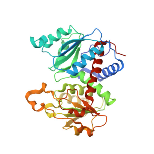
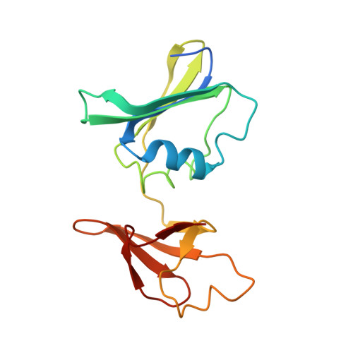
The Pathway of Product Release from the R State of Aspartate Transcarbamoylase.

Mendes, K.R.Kantrowitz, E.R.

(2010) J Mol Biology 401: 940-948

  • DOI: https://doi.org/10.1016/j.jmb.2010.07.003
  • Primary Citation Related Structures: 
    3MPU

  • PubMed Abstract: 

    The pathway of product release from the R state of aspartate transcarbamoylase (ATCase; EC 2.1.3.2, aspartate carbamoyltransferase) has been determined here by solving the crystal structure of Escherichia coli ATCase locked in the R quaternary structure by specific introduction of disulfide bonds. ATCase displays ordered substrate binding and product release, remaining in the R state until substrates are exhausted. The structure reported here represents ATCase in the R state bound to the final product molecule, phosphate. This structure has been difficult to obtain previously because the enzyme relaxes back to the T state after the substrates are exhausted. Hence, cocrystallizing the wild-type enzyme with phosphate results in a T-state structure. In this structure of the enzyme trapped in the R state with specific disulfide bonds, we observe two phosphate molecules per active site. The position of the first phosphate corresponds to the position of the phosphate of carbamoyl phosphate (CP) and the position of the phosphonate of N-phosphonacetyl-l-aspartate. However, the second, more weakly bound phosphate is bound in a positively charged pocket that is more accessible to the surface than the other phosphate. The second phosphate appears to be on the path that phosphate would have to take to exit the active site. Our results suggest that phosphate dissociation and CP binding can occur simultaneously and that the dissociation of phosphate may actually promote the binding of CP for more efficient catalysis.


  • Organizational Affiliation
    • Department of Chemistry, Boston College, Merkert Chemistry Center, Chestnut Hill, MA 02467-3807, USA.

Macromolecule Content 

  • Total Structure Weight: 155.21 kDa 
  • Atom Count: 11,363 
  • Modeled Residue Count: 1,359 
  • Deposited Residue Count: 1,389 
  • Unique protein chains: 2

Macromolecules

Find similar proteins by:|  3D Structure
Entity ID: 1
MoleculeChains  Sequence LengthOrganismDetailsImage
Aspartate carbamoyltransferase catalytic chain
A, C, E
310Escherichia coli K-12Mutation(s): 2 
Gene Names: b4245JW4204pyrBpyrI
EC: 2.1.3.2
UniProt
Find proteins for P0A786 (Escherichia coli (strain K12))
Explore P0A786 
Go to UniProtKB:  P0A786
Entity Groups
Sequence Clusters30% Identity50% Identity70% Identity90% Identity95% Identity100% Identity
UniProt GroupP0A786
Sequence Annotations
Expand
Reference Sequence
Find similar proteins by:|  3D Structure
Entity ID: 2
MoleculeChains  Sequence LengthOrganismDetailsImage
Aspartate carbamoyltransferase regulatory chain
B, D, F
153Escherichia coli K-12Mutation(s): 0 
Gene Names: pyrIb4244JW4203
UniProt
Find proteins for P0A7F3 (Escherichia coli (strain K12))
Explore P0A7F3 
Go to UniProtKB:  P0A7F3
Entity Groups
Sequence Clusters30% Identity50% Identity70% Identity90% Identity95% Identity100% Identity
UniProt GroupP0A7F3
Sequence Annotations
Expand
Reference Sequence

Small Molecules

Ligands 2 Unique
IDChains Name / Formula / InChI Key2D Diagram3D Interactions
PO4

Query on PO4


Download:Ideal Coordinates CCD File
G [auth A]
H [auth A]
J [auth C]
K [auth C]
M [auth E]
G [auth A],
H [auth A],
J [auth C],
K [auth C],
M [auth E],
N [auth E]
PHOSPHATE ION
O4 P
NBIIXXVUZAFLBC-UHFFFAOYSA-K
ZN

Query on ZN


Download:Ideal Coordinates CCD File
I [auth B],
L [auth D],
O [auth F]
ZINC ION
Zn
PTFCDOFLOPIGGS-UHFFFAOYSA-N

Experimental Data & Validation

Experimental Data

  • Method: X-RAY DIFFRACTION
  • Resolution: 2.85 Å
  • R-Value Free:  0.240 (Depositor), 0.239 (DCC) 
  • R-Value Work:  0.173 (Depositor), 0.173 (DCC) 
  • R-Value Observed: 0.177 (Depositor) 
Space Group: H 3 2
Unit Cell:
Length ( Å )Angle ( ˚ )
a = 120.711α = 90
b = 120.711β = 90
c = 692.471γ = 120
Software Package:
Software NamePurpose
ADSCdata collection
PHENIXmodel building
PHENIXrefinement
HKL-2000data reduction
HKL-2000data scaling
PHENIXphasing

Structure Validation

View Full Validation Report


Entry History 

Deposition Data

Revision History  (Full details and data files)

  • Version 1.0: 2010-07-21
    Type: Initial release
  • Version 1.1: 2011-07-13
    Changes: Version format compliance
  • Version 1.2: 2023-09-06
    Changes: Data collection, Database references, Derived calculations, Refinement description
  • Version 1.3: 2024-11-20
    Changes: Structure summary