3MPH | pdb_00003mph

The structure of human diamine oxidase complexed with an inhibitor aminoguanidine


Experimental Data Snapshot

  • Method: X-RAY DIFFRACTION
  • Resolution: 2.05 Å
  • R-Value Free: 
    0.215 (Depositor), 0.218 (DCC) 
  • R-Value Work: 
    0.177 (Depositor), 0.182 (DCC) 
  • R-Value Observed: 
    0.179 (Depositor) 

Starting Model: experimental
View more details

wwPDB Validation 3D Report Full Report

Validation slider image for 3MPH

This is version 2.1 of the entry. See complete history

Literature

Correlation of active site metal content in human diamine oxidase with trihydroxyphenylalanine quinone cofactor biogenesis

McGrath, A.P.Caradoc-Davies, T.Collyer, C.A.Guss, J.M.

(2010) Biochemistry 49: 8316-8324

  • DOI: https://doi.org/10.1021/bi1010915
  • Primary Citation Related Structures: 
    3MPH

  • PubMed Abstract: 

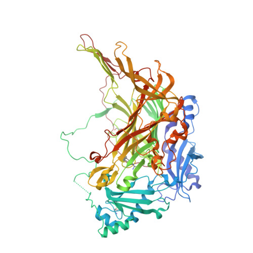
    Copper-containing amine oxidases (CAOs) require a protein-derived topaquinone cofactor (TPQ) for activity. TPQ biogenesis is a self-processing reaction requiring the presence of copper and molecular oxygen. Recombinant human diamine oxidase (hDAO) was heterologously expressed in Drosophila S2 cells, and analysis indicates that the purified hDAO contains substoichiometric amounts of copper and TPQ. The crystal structure of a complex of an inhibitor, aminoguanidine, and hDAO at 2.05 Å resolution shows that the aminoguanidine forms a covalent adduct with the TPQ and that the site is ∼75% occupied. Aminoguanidine is a potent inhibitor of hDAO with an IC(50) of 153 ± 9 nM. The structure indicates that the catalytic metal site, normally occupied by copper, is fully occupied. X-ray diffraction data recorded below the copper edge, between the copper and zinc edges, and above the zinc edge have been used to show that the metal site is occupied approximately 75% by copper and 25% by zinc and the formation of the TPQ cofactor is correlated with copper occupancy.


  • Organizational Affiliation
    • School of Molecular Bioscience, University of Sydney, Sydney, NSW 2006, Australia.

Macromolecule Content 

  • Total Structure Weight: 170.04 kDa 
  • Atom Count: 12,719 
  • Modeled Residue Count: 1,424 
  • Deposited Residue Count: 1,462 
  • Unique protein chains: 1

Macromolecules

Find similar proteins by:|  3D Structure
Entity ID: 1
MoleculeChains  Sequence LengthOrganismDetailsImage
Amiloride-sensitive amine oxidase
A, B
731Homo sapiensMutation(s): 1 
Gene Names: ABP1
EC: 1.4.3.22
UniProt & NIH Common Fund Data Resources
Find proteins for P19801 (Homo sapiens)
Explore P19801 
Go to UniProtKB:  P19801
PHAROS:  P19801
GTEx:  ENSG00000002726 
Entity Groups
Sequence Clusters30% Identity50% Identity70% Identity90% Identity95% Identity100% Identity
UniProt GroupP19801
Glycosylation
Glycosylation Sites: 3Go to GlyGen: P19801-1
Sequence Annotations
Expand
Reference Sequence

Oligosaccharides

Help  
Entity ID: 2
MoleculeChains Length2D Diagram GlycosylationD Interactions
beta-D-mannopyranose-(1-4)-2-acetamido-2-deoxy-beta-D-glucopyranose-(1-4)-2-acetamido-2-deoxy-beta-D-glucopyranose
C
3N-Glycosylation
Glycosylation Resources
GlyTouCan: G15407YE
GlyCosmos: G15407YE
GlyGen: G15407YE
Entity ID: 3
MoleculeChains Length2D Diagram GlycosylationD Interactions
2-acetamido-2-deoxy-beta-D-glucopyranose-(1-4)-2-acetamido-2-deoxy-beta-D-glucopyranose
D, E, F, G, H
2N-Glycosylation
Glycosylation Resources
GlyTouCan: G42666HT
GlyCosmos: G42666HT
GlyGen: G42666HT

Experimental Data & Validation

Experimental Data

  • Method: X-RAY DIFFRACTION
  • Resolution: 2.05 Å
  • R-Value Free:  0.215 (Depositor), 0.218 (DCC) 
  • R-Value Work:  0.177 (Depositor), 0.182 (DCC) 
  • R-Value Observed: 0.179 (Depositor) 
Space Group: P 21 21 21
Unit Cell:
Length ( Å )Angle ( ˚ )
a = 92.685α = 90
b = 94.604β = 90
c = 196.051γ = 90
Software Package:
Software NamePurpose
SCALAdata scaling
REFMACrefinement
PDB_EXTRACTdata extraction
MOSFLMdata reduction
REFMACphasing

Structure Validation

View Full Validation Report


Entry History 

Deposition Data

Revision History  (Full details and data files)

  • Version 1.0: 2010-09-08
    Type: Initial release
  • Version 1.1: 2011-07-13
    Changes: Version format compliance
  • Version 1.2: 2012-04-18
    Changes: Derived calculations
  • Version 1.3: 2014-03-05
    Changes: Database references
  • Version 2.0: 2020-07-29
    Type: Remediation
    Reason: Carbohydrate remediation
    Changes: Atomic model, Data collection, Database references, Derived calculations, Structure summary
  • Version 2.1: 2023-11-01
    Changes: Data collection, Database references, Refinement description, Structure summary