


Structural basis of resveratrol regulation of myosin activity.
Schneider, J., Taft, M., Backhaus, A., Baruch, P., Fedorov, R., Manstein, D.J.To be published.
Experimental Data Snapshot
Starting Model: experimental
View more details
Entity ID: 1 | |||||
|---|---|---|---|---|---|
| Molecule | Chains | Sequence Length | Organism | Details | Image |
| Myosin-2 heavy chain | 788 | Dictyostelium discoideum AX4 | Mutation(s): 0 Gene Names: mhcA, DDB_G0286355 EC: 3.6.4.1 | 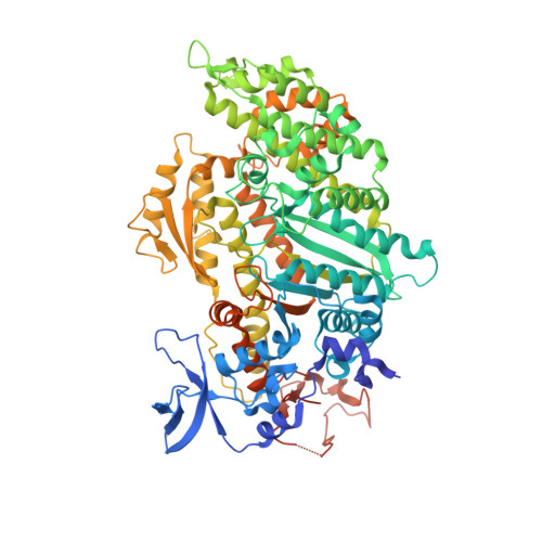 | |
UniProt | |||||
Entity Groups | |||||
| Sequence Clusters | 30% Identity50% Identity70% Identity90% Identity95% Identity100% Identity | ||||
| UniProt Group | P08799 | ||||
Sequence AnnotationsExpand | |||||
Reference Sequence | |||||
| Ligands 4 Unique | |||||
|---|---|---|---|---|---|
| ID | Chains | Name / Formula / InChI Key | 2D Diagram | 3D Interactions | |
| AD9 Download:Ideal Coordinates CCD File | B [auth A] | ADP METAVANADATE C10 H16 N5 O13 P2 V XLVFTLJPBLXCED-KWIZKVQNSA-K |  | ||
| STL Download:Ideal Coordinates CCD File | D [auth A] | RESVERATROL C14 H12 O3 LUKBXSAWLPMMSZ-OWOJBTEDSA-N |  | ||
| EDO Download:Ideal Coordinates CCD File | E [auth A], F [auth A], G [auth A], H [auth A] | 1,2-ETHANEDIOL C2 H6 O2 LYCAIKOWRPUZTN-UHFFFAOYSA-N |  | ||
| MG Download:Ideal Coordinates CCD File | C [auth A] | MAGNESIUM ION Mg JLVVSXFLKOJNIY-UHFFFAOYSA-N |  | ||
| Length ( Å ) | Angle ( ˚ ) |
|---|---|
| a = 88.9 | α = 90 |
| b = 147.9 | β = 90 |
| c = 153 | γ = 90 |
| Software Name | Purpose |
|---|---|
| MxCuBE | data collection |
| CNS | refinement |
| XDS | data reduction |
| XSCALE | data scaling |
| CNS | phasing |


